What kind of pathology is this
A hernia is a protrusion of internal organs, most often the intestines, through an opening formed in the connective tissues. If the problem is not dealt with in a timely manner, the hernia may begin to increase in size due to further protrusion of intestinal loops and organs.
This can cause strangulation, poor circulation and the formation of intestinal obstruction. Such conditions not only cause severe pain and discomfort to the animal, but in a neglected state can cause the death of young animals.
Causes of the disease
A hernia can be of the following types:
- Congenital. It occurs due to a hereditary tendency to muscle weakness and low tissue elasticity. Also, the cause of this type of hernia is the expansion of the umbilical ring.
- Acquired. This pathology develops due to trauma, for example, a blow to the abdomen, a severe bruise from a fall, as well as due to the entry of pathogens into an open wound formed during cutting of the umbilical cord.
If the abdominal wall is weak, protrusion of organs and parts of the intestine will continue due to internal pressure, so this pathology absolutely cannot be ignored.
Symptoms of the disease
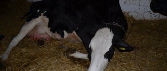
In the initial stages of the development of the disease, its signs may not be noticeable, since at this time the animal still feels well and does not suffer from pain. But a hernia in calves can be noticeable to the naked eye, so veterinarians and owners need to carefully examine the litter immediately after birth, and then regularly during the first weeks and months of the animal's life.
Expert opinion
Zarechny Maxim Valerievich
Agronomist with 12 years of experience. Our best country expert.
Ask a Question
A hernia in young animals is defined as a pineal-shaped protrusion in the navel area, soft, quite mobile, and not always painful.
In the initial stages, it can be reduced with light finger pressure, but with the slightest effort or movement of the calf, the hernia comes out again. If the problem is in an advanced stage, part of the intestine, and sometimes other organs, gets into the umbilical opening. This leads to the following symptoms:
- Pain, especially when moving or touching the affected area.
- Slight increase in temperature.
- Loss of appetite.
- Disorders of excretory function.
- Anxiety, restlessness, or lethargy in the calf.
Such signs cannot be ignored, because at any moment the hernia can be strangulated, and this poses an immediate threat to life.
See also
Why does a calf urinate red and pee in blood, what to doRead
How to properly treat an umbilical hernia in calves
If the hernia is small in size (up to 30 millimeters in diameter), it is considered non-life threatening. The animal must be monitored. Most often, nothing will have to be done, since within a year of the young animal’s life the hernia can disappear on its own due to the strengthening of the muscle corset and the closure of the umbilical opening.
In all other cases, mandatory treatment will be required, which is performed in various ways.
Conservative treatment
If a hernia in calves is detected in a timely manner, when it is still minor, there is no strangulation, inflammation or adhesions, the babies can be helped by reduction.
To do this, the veterinarian gently massages the hernia area, stimulating relaxation of the muscle ring. Then he delicately, without extra effort, pushes the tissues that have come out into the hole. To prevent repeated protrusion outward, the hernia site is sealed with a thick plaster and additionally secured with a special bandage. In order for such treatment to proceed without complications and bring relief, the calf needs to be rested for about a week.
In the future, the injured animal must be protected from physical stress, shock and injury.
Surgical intervention
If the calf's hernia is inflamed, there is suppuration, a loop of intestine or other tissue has entered the hole, there is strangulation, adhesions that threaten necrosis, urgent surgical intervention will be required.
The operation is performed by a veterinarian under local anesthesia. The navel area is cleaned of fur, disinfected and anesthetized. An incision in the abdominal wall is made 20 millimeters from the edge of the hernia. The resulting sac is removed, if necessary, the inflamed tissue is cleaned, the protruding organs are carefully adjusted, and the hole is sutured. To prevent the recurrence of a hernia in calves, fixing staples are applied to the operation site.
Postoperative period
After the intervention, the animal is provided with rest, covered with clean straw, given light, easily digestible food and constant access to clean drinking water. The calf is given antibiotics if necessary, as well as painkillers if necessary. On the fifth or tenth day after the operation, the sutures are removed, but the condition of the calf continues to be monitored. If his temperature rises, discharge or pus appears, the stitches come apart, it is necessary to take immediate action and call a veterinarian, as dangerous complications are possible.
See also
Symptoms and diagnosis of cowpox, cattle treatment and preventionRead
Help with hands and scalpel
Conventionally, types of hernias are divided into those that are simply reduced by hand and those that are removed surgically. Treatment of the former is simpler, but it is only applicable if the hernia is not too large.
First, a massage is performed, then the prolapsed part of the peritoneum is simply inserted back into the umbilical ring and sealed with an adhesive plaster. First, the hernia site is freed from hair and disinfected with alcohol. Then a special bandage is put on, and a calm environment is created for the animal. The prolapsed part of the peritoneum is eventually fixed in this position and the disease goes away.
Surgery should be performed only in a hospital setting, where the necessary equipment is available and sterility is ensured.
Local anesthesia is usually used by injecting the calf with novocaine or a similar agent. The doctor calculates the dose based on the weight of the animal.
Consequences of lack of treatment
Umbilical hernia in calves is a life-threatening pathology that requires careful attention, especially if it is strangulated. Trying to cope with this condition on your own is extremely dangerous, since adhesions may form if pinched. If you try to straighten the intestine with force, this can lead to ruptures and spillage of contents into the abdominal cavity. This threatens with an extremely serious consequence - the development of peritonitis, in which there is a high risk of losing the calf.
If hernias in calves are not treated promptly, this can cause the following complications:
- Incarceration leading to the development of necrosis and tissue rupture.
- The formation of adhesions precluding the possibility of non-surgical reduction of a hernia in a calf.
- Inflammatory process (phlegmon) of the hernial sac. It can spread to the abdominal wall and move to the affected part of the organs, accompanied by an increase in temperature and a sharp deterioration in the general well-being of the animal. The condition threatens the rapid spread of infection, damage to vital organs, general intoxication and the development of sepsis (blood poisoning).
It is impossible to assume that any hernia in calves will go away on its own. If it is detected, the animal must be under the supervision of a veterinarian.
Hernia of the white line of the abdomen: all about symptoms and treatment of pathology
A hernia of the linea alba is a pathology in which internal organs fall out of the abdominal cavity through defects in the aponeurosis, forming a hernial sac under the skin. The problem is more common in men of all ages. In women, the pathology occurs less frequently, mainly during or after pregnancy, and also at the age of 50 years. A hernia of the linea alba can also form in a child, regardless of gender. The peak of clinical cases in children occurs at 5-7 years of age.
The pathology threatens with serious complications, so we will figure out what symptoms a hernia of the linea alba appears in patients of different groups, consider all treatment methods, including non-traditional ones, and explain what disadvantages they have.
Classification of hernias of the white line of the abdomen
The linea alba is the fusion of the aponeuroses of the muscles of the anterior abdominal wall. Aponeuroses are tendon plates consisting of connective tissue collagen. The main purpose of the linea alba is to connect the left and right rectus abdominal muscles. When the integrity of the connective tissue is violated, conditions for the formation of hernias appear. In surgical practice, three types of these pathologies are distinguished.
Our readers successfully use SustaLife to treat joints. Seeing how popular this product is, we decided to bring it to your attention. Read more here...
- Supraumbilical or epigastric. Due to the weakness of the connective tissue in the area from the navel to the solar plexus, this type of hernia occurs most often.
- Paraumbilical or paraumbilical. Less common, it is often diagnosed as an umbilical hernia.
- Subumbilical. In the lower abdomen, the linea alba has the greatest strength, so this type of hernia is extremely rare in medical practice.
In especially severe cases, hernias may appear in several places, one above the other.
In addition to classification by localization of pathology, hernias are also grouped by stages. There are only three of them (see description in the table).
Note that the symptoms of a hernia at different stages may differ from those shown in the table. This depends on the location of the pathology, the contents of the hernial sac and other factors.
According to the International Classification of Diseases as amended in 2010 (ICD-10), recommended by WHO, hernia of the white line of the abdomen can be assigned to one of several groups:
A more precise code depends on the location of the pathology, the presence of obstruction or gangrene and is assigned after a diagnostic examination.
Read also: How to make ventilation in the cellar
Reasons for the development of pathology
The main reason for the formation of hernias of the white line of the abdomen is weakening or disruption of the integrity of the connective tissue due to the following factors:
- various injuries;
- lack or insufficient physical activity, including due to diseases of the musculoskeletal system, for example, synoviomas;
- obesity;
- increased intraperitoneal pressure;
- surgery (the resulting hernia is a postoperative complication).
These are the main problems that cause pathology in people regardless of age. In addition, in different gender and age groups of patients, a hernia of the white line of the abdomen can be provoked by specific causes.
Causes of hernia of the white line of the abdomen in men
In 60% of clinical cases, a hernia of the white line of the abdomen is diagnosed in representatives of the stronger sex. This is usually associated with the professional activities or hobbies of men. An epigastric hernia can occur in the following cases:
- sudden or improper lifting of weights during work or sports;
- weakening of the aponeurosis due to sedentary work (for drivers and men working in offices);
- absence of a bandage during loading and unloading operations.
Most men are careless about their health, so their hernia of the white line of the abdomen is detected in the second or third stage.
Prevention
If the formation of an umbilical hernia in offspring is associated with heredity, it is impossible to prevent it, but there is a chance to cope with it in the early stages. In other situations, you can protect the animal from the formation of a hernial sac by keeping the calf in comfortable conditions, avoiding injury, blows, and falls.
Scientists from the USA believe that a risk factor is an infection that gets into the umbilical wound, so frequent treatment with available antiseptics, for example, tincture of iodine, can be a method of prevention. This will help speed up healing and reduce the risk.
Another preventative method is the use of plastic clamps to prevent the expansion of the umbilical ring and the formation of a hernia in calves.
This condition is common in newborns and toddlers, but requires a competent approach to avoid painful and risky consequences. If you do not pay attention to the presence of a hernia, this will cause the gradual development of a hernia and inflammation, which can lead to sudden complications and death of the young animal.
What causes a hernia
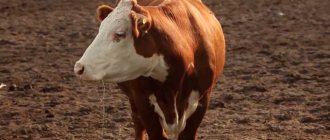
Hernia in calves is considered a non-contagious disease. This means that a sick animal is not dangerous to its fellow tribesmen and people. This does not diminish the danger of the disease, which can be congenital or acquired.
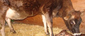
The main symptom is swelling in the abdomen at the site of the navel. When palpating this area, the baby does not experience pain, but bowel sounds are heard through it. The bulge itself is soft, and with more careful pressure, the hole under it is determined. It is oval in shape or slightly narrowed, like a slit. This formation is called a hernial ring. Part of the peritoneum protrudes from it, and in especially severe cases, internal organs.
Read also: Use of hawthorn: benefits and harm to the body
The cause of such a defect in a newborn calf is a poorly processed, and therefore long-non-healing, umbilical cord. And the causes of an acquired hernia can be various injuries - a fall of an animal, a blow from a horn, a hoof, or any hard object.