Preparing the animal
Blood is taken from cows to study its biochemical composition, exclude infectious diseases or confirm the suspected diagnosis. Venous blood is necessary for testing for leukemia, brucellosis, and tuberculosis. To take material from a cow without causing harm to the animal’s health, it is necessary to properly prepare for the procedure. The optimal time for collection is the morning hours before the first feeding. After eating, it is not recommended to take blood for analysis within 5 hours.
The site for taking the sample is cleared of hair and, if necessary, the animal is fixed, after which the area is treated with an antiseptic. Suitable for disinfection are 70% ethyl alcohol solution, 1% alcohol solution of salicylic acid, 5% alcohol solution of iodine.
Techniques that do not require forced fixation of body position are easier to tolerate by animals. Stress from the procedure can lead to a decrease in milk yield. It is not recommended to take blood from females 3 weeks before calving and within 3 weeks after birth.
Expert opinion
Zarechny Maxim Valerievich
Agronomist with 12 years of experience. Our best country expert.
Ask a Question
To avoid complications, it is necessary to follow the blood collection technique, choosing an option that is less traumatic for the cow and safe for humans.
Methods for collecting blood from cows
Today, there are several methods for collecting blood from cattle. It is taken from the following veins:
- jugular;
- dairy;
- tail vein.
Before carrying out the procedure, it is recommended to first secure the animal, which will prevent injury. In this state, the cow will also not be able to knock over the test tube. Before the procedure, you will need to disinfect the blood collection site using a solution of phenol, alcohol or iodine.
Taking a sample from the jugular vein is one of the most popular methods. As a rule, the procedure is carried out early in the morning or before the cow receives food. To carry out the procedure, the animal's head is tied and fixed in a motionless state. The needle must be inserted at an acute angle, with the tip directed towards the head.
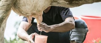
It is allowed to draw blood from the mammary vein for research only from an adult. The milk veins are located on the side of the udder and extend down the abdominal area. Through them, the mammary glands are supplied with blood and nutrients. It is worth noting that the better developed the milk veins, the more milk can be obtained from the cow.
It is safest to take samples for testing from the tail vein. The injection site, as in other cases, must be disinfected. If you choose the injection site at the level of 2 to 5 vertebrae, the procedure will go more smoothly.
Taking blood from cows from the tail vein
As practice shows, taking blood from the tail vein for research is the safest option. For these purposes, you can use a regular needle or use a special vacuum system. Such systems already include special tubes that contain an anticoagulant and the required pressure, which allows blood from the tail vein to flow smoothly into the container.
Before taking a sample from the tail vein, it is necessary to disinfect the injection site with alcohol or iodine solution. After this, the cow’s tail is lifted and held by the middle third. In this case, the needle must be inserted smoothly into the tail vein, the angle of inclination should be 90 degrees. The needle is usually inserted all the way.
This method of collection has many advantages:
- the sample taken is completely sterile;
- practically no clots form in the test tube, as a result of which all samples are suitable for research;
- This procedure does not require much time. An experienced veterinarian can call samples from 200 animals in 60 minutes;
- when using this method, there are no side effects, and the chance of injury to cattle is minimized;
- contact with blood is minimal;
- the animal does not experience stress, the usual level of milk yield is maintained.
This method is most often used on large farms where it is necessary to take a large number of samples in a short period of time.
Taking blood from cattle from the jugular vein
If it is necessary to draw blood from the jugular vein, it is recommended to insert a needle at the border where the upper third of the neck transitions to the middle third. The first step is to induce a sufficient level of filling of the vein and minimize its mobility. For these purposes, it is recommended to compress the vein using a rubber band or fingers.
During the puncture, you will need to hold the syringe with the needle in your hand so that the direction of the needle coincides with the line of the vein being punctured. It is necessary to ensure that the cut of the needle is directed upward, towards the head. The needle should be inserted at an angle of 20 to 30 degrees. If the needle gets into a vein, blood will begin to flow out of it.
Before removing the needle from the cow's jugular vein, you should first remove the rubber tourniquet and pinch the vein with your fingers. It is necessary to pinch just above the place where the needle is located. The needle is gradually removed, and it is recommended to compress the injection site with a cotton swab for a while, which will prevent the formation of hematomas on the animal’s body. At the end of the procedure, the venipuncture site is disinfected with alcohol or tincture of iodine and treated with Collodion solution.
Attention! Depending on the task at hand, blood, plasma or serum can be used for research.
Taking blood from the mammary vein
In this case, it is necessary to take into account that blood sampling from the mammary gland can only be done in adults. The required vein can be found on the side of the udder.
Before taking a sample, it is recommended to first secure the animal. As a rule, the procedure will require the presence of several people. The first step is to shave or cut off the hair from the place where you plan to puncture with a needle. After this, the prepared area is disinfected using alcohol or iodine solution.
There should be a kind of small tubercle in good visibility, where it is recommended to insert the needle. Since it is quite easy to harm a cow, the needle is inserted as carefully as possible. It must be inserted at an angle, parallel to the course of the vein, until the needle precisely hits it and dark-colored venous blood appears.
This method has some advantages:
- reasonable cost of materials required for research;
- It does not take much time to collect samples;
- minimal blood spatter.
Despite this, there are also significant disadvantages:
- the risk of injury to the cow is quite high;
- will have to come into contact with the blood of the animal;
- during blood sampling, the animal experiences severe stress, as the needle is inserted into the most tender place on the body;
- This procedure is quite difficult to carry out.
Thanks to new technologies, this method has become outdated and is practically not used in research.
Technique for collecting blood from cattle
Blood is collected from cows from the jugular, caudal or mammary veins. Work in each zone has its own characteristics, due to the different location and speed of blood flow.
From the jugular vein
In accordance with the common and well-established method of taking blood from cows from the jugular vein, a bloodletting needle and a sterile tube are used, into which liquid is drawn along the wall. The vessel is located in the lower third of the animal's neck. The head must be fixed, which becomes a stressful factor for the cow.
See also
The structure of the cow's skull and its component parts, the anatomy of a horned animalRead
Technique for collecting blood from the jugular vein:
- The animal's head is fixed in a stationary position.
- Prepare the lower third of the cervical area, removing excess hair, and disinfect the surface of the skin with an alcohol solution.
- Press the vessel with your thumb.
- The needle is inserted into the vein at an acute angle to the surface towards the head. The insertion depth is 1 centimeter.
- Collect blood in a test tube.
Material collected in this way is not sterile and liquid may splash.
From the milk vein
The milk vein is located on the cow's abdomen on both sides, on the side of the udder. It is clearly visible in adult females, but the sampling process is complicated by the high sensitivity of this part of the body and the deeper location of the vein than it seems visually.
The cow must be securely secured and held, which often requires more than one person.
Technique for collecting blood from the mammary vein:
- The animal is secured and restrained.
- Remove hair from the area adjacent to the vein.
- Treat the area with an alcohol solution.
- Feel the venous tubercle with your fingers.
- The needle is inserted into the vessel parallel to the surface of the skin.
- Collecting biomaterial.
The procedure is unpleasant for the cow and can cause a decrease in milk yield as a result of the resulting stress. This method, complex and traumatic for the animal, is rarely used in modern conditions.
From the tail vein
Taking blood from the tail vein is quick, does not require forced restraint of the animal, and is usually easily tolerated by the animal. Modern techniques are aimed at developing devices for collecting material from this particular area of the cow’s body.
Rules for taking blood from the tail vein:
- Take the cow's tail in the middle of its length with your hand and lift it up.
- Disinfect the area of 2-5 vertebrae and adjacent areas with an alcohol solution.
- Take a sterile needle or a ready-made special system in one hand, and hold the tail with the other.
- The needle is inserted perpendicular to the center of the width of the tail at a distance of about 10 centimeters from its base. The insertion depth is 0.5-1 centimeter.
- Collecting material.
See also
How to properly feed a cow at home before and after calvingRead
The procedure eliminates human contact with biological fluids of livestock, which is considered one of the main advantages. Slow blood flow in the vessel creates difficulties during sampling, but modern vacuum systems solve this problem, making the procedure safe and effective.
Preparing for blood collection from cattle
As a rule, blood is drawn from cows from the jugular vein, located in the upper third of the neck. The volume of material obtained for research should not be less than 5 ml with an anticoagulant of 0.5 M EDTA.
Before starting the procedure, the needles used should be pre-sterilized using boiling for this purpose. It is important to consider that sampling from each cow must be done with a new needle.
The collection site must be disinfected. For disinfection, use alcohol or a 5% iodine solution. During sampling, the animal must be securely restrained - the head is tied.
After the material for the study has been taken, it is worth tightly closing the tube and turning it over several times so that mixing with the anticoagulant occurs. In this case, shaking is not allowed. Each test tube is numbered according to the inventory.
The most effective way is to take blood from the subcaudal vein. In this case, it is not necessary to restrain the cow. It is recommended to further store the tubes at a temperature range from +4°C to +8°C. A refrigerator is perfect for these purposes. Do not use the freezer. If clots appear in the sample taken, it is unsuitable for further research.
Attention! The use of heparin and other types of anticoagulants is not allowed. To transport the intake material, special bags with refrigerant are used. During transportation, blood should not be clotted or frozen.
Features of vacuum blood sampling
The use of modern vacuum systems for blood collection allows the KRS procedure to be carried out quickly and safely. The vacuum method works well on the tail vein. The process does not injure the cow and eliminates direct contact of humans and other animals with the biomaterial.
The system consists of a needle and a syringe container. The needle, which has an optimal diameter (usually 0.9 millimeters), is equipped with a valve that prevents fluid from leaking out, which reduces the likelihood of possible complications. The syringe, which also functions as a transport container, is made of durable plastic. You can also isolate serum or add an anticoagulant.
Advantages of the vacuum method:
- no need to restrain the animal;
- minimizing the stress factor for the cow;
- exclusion of direct human contact with the material;
- obtaining a sterile sample;
- eliminating the unforeseen risk of spreading infection;
- ease of use without transferring the sample into a transport container.
Marking of vacuum systems includes practical color coding, which allows you to sort the samples taken.
Taking blood from pigs
The best place for the procedure is the vessels of the ears, the tip of the tail, the ophthalmic sinus, and the anterior vena cava.
Many specialists cut off the tip of the tail, which leads to hemolysis. At the same time, the sterility of the sample leaves much to be desired. The picture is worsened by bleeding and resulting cannibalism.
It is more effective to take material from the anterior vena cava, but this requires some experience. In addition, this method is labor-intensive.
It is more convenient to take the material from the ophthalmic sinus. Biosample collection strategy:
- The pig is secured by the upper jaw using a loop. Piglets can be restrained in a dorsal position, and head movement can be further limited by hand.
- Blood is drawn using a vacuum syringe. It is allowed to use an ordinary needle and test tube.
- The needle is inserted into the inner corner of the eye under the third eyelid, beveled towards the bone (not towards the eyeball!). At the same time, the cannula is manipulated so as to push the eyeball away with the needle (conditionally focusing the tip on the opposite ulnar tubercle).
With experience, the required depth of needle insertion is easily determined: 2-3 cm in adult pigs, 0.5-1.0 cm in piglets. Beginning veterinarians can insert the needle until they feel it touch the bone.
- If everything is done correctly, then when the needle is removed from the bone, the blood should begin to flow on its own. As a rule, a 20 ml tube is filled in one and a half minutes (faster if the animal squeals).
- Dripping blood from the puncture site is removed with a swab.
Possible mistakes
When performing the standard technique of taking blood from the jugular vein, there is a high probability of the material getting into contact with a person and surrounding objects. If an animal is infected, there is a risk of spreading the infection. The open method takes a lot of time, requires preparation and extreme care.
If the rules of asepsis and blood sampling techniques are violated, the resulting sample may not meet the requirements, and the cow may experience complications in the form of abscesses and hematomas. It is not recommended to force the collection to avoid partial hemolysis of blood cells.
blood collection using vacuum tubes
Dear forum users! Tell me if there are materials or methodological recommendations on how to collect blood from cattle from the tail vein using vacuum tubes. Because I bought them, test tubes, but our veterinarians are not trained to take blood. All suppliers say that it is convenient and fast, but the training materials are only for people.
One year I took blood with such test tubes, it’s very convenient, you insert the needle into the middle of the tail vertebra from below, then you insert the test tube, the vacuum sucks in the blood, that’s it, the cow doesn’t twitch, you take it quickly, beauty, try two or three, then it goes like clockwork, I also started by trial and error no one knew how to take mistakes, and then they said it was expensive and the old fashioned way with a needle through the jugular vein.
Symptoms of brucellosis in cows
The main and most obvious symptoms of brucellosis in cows are:
- abortions;
- birth of a dead calf.
In some cases, an external symptom may be the formation of bursitis on the cow’s legs, as well as abscess inflammation and hygroma on the hind legs. Cattle may experience loss of appetite, decreased activity, and sometimes the temperature may rise.
When the bacterium settles in the animal’s body, over time the disease begins to progress in the internal organs: liver, spleen, mammary glands and uterus.
Brucellosis is a latent disease that occurs without any symptoms. The absence of symptoms makes it extremely difficult to identify the disease in the early stages of development.
It is important to know that abortion is not always a symptom of the disease. Sometimes the calf survives, but after birth it is very weak.
We recommend reading: Diagnosis and treatment of autoimmune skin diseases in dogs and cats