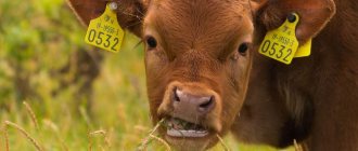
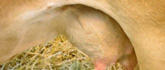
What is beef tripe?
The outwardly unattractive and, to be honest, not very pleasant-smelling section of the cow's stomach is a large muscular sac. It pre-digests plant foods - fresh grass, dry hay. It is elastic and holds up to 50 kg of feed.
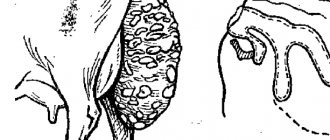
Slightly slimy to the touch and unsightly in color, the vital organ of the cattle digestive system is covered in numerous growths inside, giving it a fur-like appearance. And with all this, it is a storehouse of useful substances, including enzymes and beneficial microorganisms that have a positive effect on the gastrointestinal tract of pets - they improve digestion and heal the intestinal microflora.
Both raw and processed tripe can be added to dogs' food. Cleaning a cow's stomach is done as follows: after washing in warm water, the offal is placed in a special drum, where, under the influence of steam and boiling water, the top layer (“fur”) is softened and cleaned off (the presence of a ribbed disk in the device facilitates this process). This treatment partially eliminates the unpleasant odor and allows you to prepare food for the dog.
But processing waste can also be used - this is an excellent dietary food for young puppies, elderly animals weakened after a serious illness or surgery.
Important!
In some national cuisines, peeled boiled tripe is considered a delicacy; such a dish was served on the rich merchant and boyar table in Rus'.
How does a scar work?
In calves, the rumen, mesh and book are not developed. One abomasum is functioning. Newborn calves do not have cud. They still have nothing to chew. All proventriculi are a single elongated tube. The tube has exits for the scar, mesh and book, but they are tightly closed with the lips. The tube is a continuation of the esophagus. The rennet is small in size, so it is important that the calf drinks the milk in small sips.
When feeding young animals from a bucket, calves can swallow large portions of liquid. The milk enters through the lips into the forestomach and begins to sour there. Gastrointestinal disorders develop. Newborn calves are fed from a teat or brought to the udder of a cow.
From 2 weeks, the young animals begin to be given hay. This is roughage. It requires the functioning of the rumen. Gradual feeding of roughage in small portions promotes its development. Hay from the rumen passes into the mesh, but the mesh does not allow it to pass further through the gastrointestinal tract. The factions are too rough.
More on the topic: Peculiarities of feeding cows after calving and during milking
The animals belch and the plant food returns to the oral cavity. In the mouth, under the influence of a large amount of saliva and when grinding with teeth, food is crushed into pulp. Calves develop chewing cud. They swallow little by little liquid gruel, which enters the mesh and passes further through the proventriculus into the intestines.
What is a scar? – This is the largest section of the stomach. In adult animals its volume is 200 liters. It occupies most of the abdominal cavity. The scar is located on the left side from the diaphragm to the pelvic cavity. If there is a need to massage the proventriculus, then approach the cow from the left side.
The scar consists of 3 chambers: dorsal, ventral and cranial. They are called bags. They are connected by longitudinal gutters. The inner part of the gutters forms muscle cords that are covered with a mucous membrane. The shell forms folds. The largest bag is the dorsal one. It is located horizontally in the abdominal cavity.
Closer to the pelvic cavity is the ventral sac. At the bottom of the cavity, vertical to the dorsal chamber, is the cranial sac. With pathology of the gastrointestinal tract, food stagnation most often occurs in it. The ventral and cranial sacs are small in size.
There are no glands in the rumen. The entire surface of the mucous membrane is covered with papillae. They increase the absorptive surface of the proventriculus. Digestion of feed in the cavity occurs under the influence of bacteria and microorganisms.
- Bacteria in the forestomach are 7 kg. They occupy 10% of the cavity. They break down starch, fats, and proteins. Stimulates the growth of bacteria alfalfa, clover, timothy.
- There are 23 species of fungi in the rumen. These are mold and yeast. They affect cellulose. Mushrooms promote the production of B vitamins, streptomycin, tetracycline, which are antibiotics.
- Microorganisms in the forestomach are 2 million/1 ml. They are involved in the digestion of roughage and dry food. Ciliates are capable of synthesizing proteins of their body. The animal assimilates proteins along with proteins that enter the body along with food. Protein produced by ciliates is considered more useful.
When feeding, it is necessary to prescribe a specific diet for cows. This will keep the number of bacteria, microorganisms and fungi in the correct ratio.
A large amount of legume feed will increase the number of bacteria, but will reduce the level of ciliates. Grain mixtures help increase the number of ciliates, but they will have a detrimental effect on the bacteria in the forestomach.
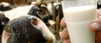
Benefits of beef tripe
A cow's or sheep's stomach contains many nutrients. It consists of three layers, but the most useful for dogs is considered to be the inner one, which consists of glands and growths (papillae). In the middle layer, consisting of muscle fibers, there are nutrients, but there are fewer of them, and the outer layer practically does not have such beneficial properties.
Lamb tripe can also be used as food for dogs, but its nutritional value is lower than that of beef tripe, which contains:
- vitamins – group B (B1, B2 and others), PP;
- microelements - phosphorus, calcium, potassium, sulfur, magnesium, sodium, iron, iodine;
- amino acids - omega-3, omega-6 and others.
The microflora of the stomach of ruminants has a beneficial effect on the dog's digestion. In the wild, wolves - the closest relatives of domestic dogs - primarily eat the victim's tripe.
Important!
Most enzymes and beneficial microorganisms are contained in unpeeled rumen; after heat treatment, some of the beneficial properties of the product are lost.
Functions of the cow's rumen
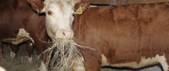
The main thing in a cattle diet is hay. Rough food forms a “cushion” when it enters the mouth. In turn, due to the contraction of the cow's rumen, it is constantly shaken. As a result, the food becomes moist, swells and grinds. After this, the animal is allowed to be given succulent and dry food.
The solids help other foods mix with bacteria. After grinding, they move more intensively to the next section - the mesh. Between it and the scar there is an exit consisting of epithelium, but without papillae.
When feeding immediately with succulent or dry food, the food moves towards the liquid contents of the rumen and then settles on the walls. The feed does not mix with each other, which is why the swollen mass does not interact with the microflora.
The bolus of food enters the mesh and then quickly moves through the proventriculus. As a result, most of the microelements are not absorbed and are excreted from the body along with feces. The optimal mode for normal decomposition is created at a temperature of 40 degrees and a pH value of 5-7.
In this case, the following happens in the forestomach:
- fiber breaks down into glucose, and starch into glycogen and amylopectin;
- non-volatile and volatile fats are formed;
- the protein is broken down into amino acids and peptides, and ammonia is also released;
- under the influence of bacteria, fats are converted into glycerides, waxes, lipids, sterols and free fatty acids;
- microflora is responsible for the synthesis of B vitamins.
Expert opinion
Kosheleva Maria Andreevna
Farmer with 15 years of experience raising goats and cows.
One part of the beneficial microelements is absorbed into the body through papillae on the rumen mucosa. The other enters the intestines and proventriculuses, after which it is distributed throughout the body through the blood.
When functioning, the rumen releases an increased amount of gases. If you do not monitor the animal’s diet, you will have to contact a veterinarian for help. The veterinarian will check the condition of the forestomach and prescribe appropriate treatment for the cow's rumen.
However, in the absence of proper therapy, serious diseases develop in the body of cattle. The most common are acidosis and rumen tympany in cows. To provide first aid, the farmer must determine where the cranial chamber is located. This is due to the fact that with the development of pathology, all gases accumulate in this place.
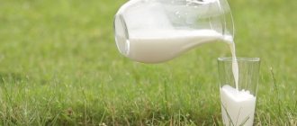
Can it be given to puppies?
Beef tripe is introduced into the menu for pets from 5-6 months of age. Since any new dish can cause a negative reaction in the body, you need to give the treat for the first time carefully - in small quantities, observing the reaction of the pet’s body for several days. You can gradually increase the number of feedings per week up to 5 times.
The norm is 100 grams per 10 kg of dog weight. Boiled tripe can replace one of the daily feedings. The unrefined raw product is given as a treat and reward in small quantities. You can alternate a raw dish with a boiled one.
Experts recommend giving elderly dogs cow by-product to maintain intestinal microflora daily, but not more than 200 g. For dogs of large (working) breeds to gain muscle mass - 100 g several times a day, for a medium-sized dog - twice a day for 100 g.
Modern view of the microbiome
Almost 90% of rumen microorganisms are uncultivable and previously unknown, and new uncultivable species were discovered within known taxa - ruminococci, eubacteria, clostridia, lactobacilli, etc.
It became clear why classical microbiological approaches previously did not make it possible to establish a statistical relationship between rumen microflora, diets, health, productivity and other indicators: not all the desired microorganisms were identified. The main inhabitants of the cattle rumen were microorganisms directly or indirectly associated with the fermentation processes of plant feed.
Average content of microorganisms in the rumen of a clinically healthy cow
First of all, these are fungi - chytridiomycetes, which are the main initiators of colonization of lignocellulosic materials. As well as methanogenic archaea, bacteria - amylolytics and cellulolytics. And lactate-utilizing bacteria that ferment a number of acids, including lactic acid. Therefore, by 2–3 months in the rumen of calves, the concentration of microorganisms that break down plant feed reaches a level characteristic of an adult animal.
It was found that the content of many microorganisms in the rumen of cattle fluctuates throughout the day. This is due to the processes of rumen fermentation: the formation of volatile fatty acids (VFA), ammonia and other substances. However, contrary to traditional opinion, in the rumen of clinically healthy animals, in addition to representatives of normal microflora, pathogens of various diseases were detected in small quantities - enterobacteria (capable of causing gastroenteritis), fusobacteria (necrobacteriosis), staphylococci and campylobacteria (mastitis), etc. This indicates about their constant presence in the rumen ecosystem of clinically healthy animals. Molecular genetic studies, in contrast to traditional cultures on nutrient media, make it possible to identify statistically significant differences in the composition of the rumen microbiome of ruminants of different ages, levels of productivity and health. For the first time in world practice, the data obtained made it possible to determine the boundaries of the normal content of microorganisms in the rumen - representatives of beneficial, undesirable and opportunistic microbiota - in accordance with the age and physiological state of the animal. These standards are widely in demand on farms today. They are used to assess the state of the rumen microbiome.
How to cook properly
If you give your dog a raw product, it should first be frozen thoroughly so that all parasites that may be in the product are killed. The offal should be kept in the freezer for at least three days, it is best to immediately divide it into portions weighing 100 g.
After freezing, the portion required for feeding should be cut into small pieces, approximately 2.5 * 2.5 cm in size, or crushed using a meat grinder. It is recommended to give tripe by adding it to porridge or mixing it with meat twice a week as a nutritional supplement.
Beef stomach can be fed boiled. To cook after defrosting, cut it into medium-sized pieces and cook for at least 3-4 hours, regularly skimming off the foam. After cooling, you can cut into smaller pieces and mix with porridge or vegetables. Many people bring the product to a boil in ordinary water, and then finish cooking it in meat broth, cooked in advance.
Important!
Raw beef gizzard should not be given to your pet daily. Feeding frequency: 2-4 times a week.
A more labor-intensive process is preparing a dried delicacy from beef tripe. You've probably seen this product prepared in a pet store, but you can prepare it yourself. Rinse and clean the raw beef gizzard well. Then cut it into even pieces and dry it in the fresh air, while covering it from direct sunlight. No need to add salt or spices!
Scar mucosa and its functions
The mucous membrane of the scar is represented by flat multilayered epithelium, slightly keratinized and forming villi, which increase the surface of the scar by approximately 7 times.
Cattle have about 520 thousand villi. About 80-85% of the entire surface of the mucous membrane is covered with villi. There are villi of different shapes - ribbon-shaped, leaf-shaped, dome-shaped, in the form of tongues, warts, etc. Their sizes range from 2 (length) x 1 mm (width) to 9 x 3 mm. In various zones of the scar, due to the formation of villi, the active surface can increase by 14-21.6 times. Often in the rumen of cattle there are villi measuring more than 12 x 5 mm. The highest density of large villi in all studied animals is observed in the vestibule of the rumen. Both species-specific differences in the structure of the relief of the rumen mucosa, as well as fundamentally similar structures independent of species, determined by the type of nutrition, are described. The relief of the mucous membrane of the rumen in wild animals feeding on roughage corresponds to that of domestic ruminants. In animals that prefer soft food (giraffe, gazelles), in all areas of the rumen the mucous membrane is densely and evenly covered with villi. The largest villi appear to be found in the giraffe rumen (22 x 7 mm).
Multilayer epithelium, 200-300 microns thick, has 15-20 rows of cells, divided into 4 layers - basal, spinous, transitional, horny. The basal layer (Str. basale) consists of a single row of cells in direct contact with the basement membrane separating the epithelium and the lamina propria (Lamina propria). Cells are adjacent to the basement membrane either by their flattened base or by long cytoplasmic processes that extend both from the base of the cell and from its lateral surfaces. The cell nuclei are round or oval in shape and are located in the lower third of the cell. There are many mitochondria in cells. The spinous layer (Str. spinosum) consists of 2-20 rows of cells of irregular polygonal shape, the highly elongated processes of which can reach the basement membrane. The spinous shape of the cells is explained by the presence of numerous short processes, with the help of which neighboring cells contact each other. The cell nuclei are round in shape, and there are fewer mitochondria than in the cells of the basal layer. As they approach the transitional layer (Str. transitionale), the epithelial cells flatten and are oriented parallel to the surface of the layer. This layer is morphologically heterogeneous and consists of 2-3 rows of highly flattened cells with folded membranes. In cell nuclei, compaction of nuclear material and shrinkage are observed. Dense fibrillar material accumulates along the cell periphery. The cells contain both larger granules and fine fibrillar and lamellar structures. The transition to the stratum corneum (Str. corneum) occurs suddenly, as a kind of “leap in keratinization.” At the same time, nuclear derivatives containing DNA are preserved in many keratinized cells. There are three types of cells. Scaly cells are especially closely connected to each other. In the scale-like horn cells, a maximum of one slit-like cavity can be found. These cells consist of a homogeneous or cellular horny substance. Spindle cells are characterized by the presence of a wide peripheral zone of keratin and an expanded intracellular space with amorphous and granular contents. The cell membranes of both cell types are highly folded. Pear-shaped cells were also noted, which are characterized by the presence of a thick keratinized wall; fibrillar material is located in the center of the large cellular space. During desquamation (desquamation), interconnected horny scales or individual horny cells are separated. At the junction of neighboring cells in the ruminal epithelium, desmosomes are formed, penetrated by tonofibrils. Cells Str. basale are connected to the basement membrane by hemidesmososmata (hemidesmosomes) In Str. spinosum and Str. transitionale forms significantly more desmosomes than in Str. basale. The dimensions of the intercellular spaces decrease as we move from Str. basale to Str. transitionale. Already in Str. basale and Str. spinosum, fusions of the outer layers of the cell membrane are found. These Macule occludentes are located in the desmosome region of two adjacent cells. On the border between Str. transitionale and Str. corneum there are elongated fusions of membranes, which, in the form of Zonulae occludentes, close the intercellular spaces. Intercellular gaps between scale-like horn cells Str. corneum are very narrow.
A detailed analysis of the ultrastructure of the epithelial layer lining the surface of the scar shows that the scar wall and especially the mucous membrane have important physiological functions, primarily in maintaining the constancy of the scar contents. Thanks to the system of endplates (Zonulae occludentes), the internal contents of the scar are reliably fenced off from the internal environment of the body, primarily from the lamina propria mucoae. It contains a powerful capillary network of the scar mucosa, the branches of which penetrate almost to the epithelium.
The mucosa has bilateral permeability, which ensures passive transport of water and ions into the blood and back according to the laws of osmosis and active transfer of substances by phago-, pinot- and exocytosis. A special role is played by the basal layer, which carries out the active transport of metabolites, primarily volatile fatty acids (VFA) and ammonia. Due to the ability to transport metabolites from the blood into the rumen cavity, the host body can influence the population of microorganisms.
The stratum corneum of the rumen epithelium acts as a reliable bacterial filter. Bacteria are found only in burst pyriform horn cells or wide intercellular spaces between these cells. Superficial layers determine the passage of water and soluble metabolites through the epithelium. If the surface of the mucosa from the side of the rumen cavity is exposed to hydrostatic pressure of the order of 20-40 cm of water. Art., then the passage of water towards the serous membrane increases. Pressure from the serous membrane causes a gradual and strong increase in the flow of water towards the cavity. Under these conditions, expansion of intercellular spaces and damage to the epithelium, expressed in the formation of vacuoles, is observed. This condition can allow water to enter the rumen and dilute its contents during acidosis.
The barrier functions of the surface layers are mainly associated with the Zonulae occludentes region. It is here that the passage of substances is difficult, if not completely impossible. It is possible that this area functions as a selective absorption filter, permeable to high-molecular substances with a particle size of 75 A. The highly branched subsystem of Zonulae occludentes tubules, formed by slit-like intercellular spaces, creates favorable conditions for the transport of substances between cells. Intracellular transport is facilitated by numerous contacts between neighboring and even very distant cells. It is assumed that in the deep layers of the rumen epithelium there is another functional barrier that limits the flow of water through the rumen wall.
The absorption, accumulation and intracellular digestion of high-molecular substances, as well as their transport through the surface layers of the rumen mucosa, are carried out by a system of phagosomes and heterolysosomes, which carry out controlled transport through the rumen epithelium. Even horn cells retain the ability to form membrane vesicles, and therefore the cells can perform such important functions as phagocytosis and exocytosis. Membrane vesicles can move inside cells, bypassing the cells of the keratin skeleton of horn cells. Diffusely distributed in Str. corneum hydrolases (esterases, acid phosphatase) begin to digest substances that end up in heterolysosomes as a result of phagocytosis.
Diffusion processes through the rumen epithelium are largely determined by higher permeability for lipophilic metabolites than for hydrophilic ones. This is explained by the fact that lipids pass more easily through the lipid regions of membranes, while hydrophilic substances must diffuse through water-filled pores. Thus, diffusion depends not only on the chemical or electrochemical gradient, but also on the physicochemical properties of the diffusing metabolite itself. Qualitative differences in the permeability of cytoplasmic membranes under conditions of unequal distribution of these parameters in the cell leaves a prerequisite for an active directed trans-aorta, which is especially important in cases where specific transporters are not involved.
This position received the following experimental confirmation. Inhibition of Na+ transport by ouabain (a specific inhibitor of Na+-, K+-ATPase) is observed only if the inhibitor acts on the serous side of the mucous membrane. In relation to blood, the contents of the rumen are electronegative, and this electrochemical potential is explained by the transport of Na+. The transepithelial potential difference increases with increasing Na concentration and disappears when transport is suppressed by ouabain or during oxygen starvation. In in vitro experiments, a maximum potential of 15 mV was recorded in the rumen of sheep, and 36 mV in calves; In vivo, the potential difference in sheep is about 30 mV. Thus, more than half of the Na from feed and saliva (in sheep 1200 mEq) is actively transported through the rumen epithelium.
Along with the mechanism of the ion pump for strong electrolytes, a nonspecifically acting pump for the active transport of weak electrolytes was also discovered in the ruminal epithelium. The driving force of such a pump is the constancy of the electrochemical potential difference of hydrogen ions between the tissue and the surrounding internal liquid media (blood, lymph). In this case, both dissociated and non-dissociated molecules can enter the epithelial cells, and only non-dissociated compounds enter the blood.
The metabolism of scar epithelium also affects passive transport carried out by diffusion. This occurs, firstly, during the transport of dissociated substances under the influence of the ruminal potential, which stimulates the diffusion of anions from the rumen into the blood and inhibits this process for cations. In accordance with the electrochemical potential difference, the diffusion of monovalent cations becomes possible when the concentration of this ion in the blood is 3-fold, and divalent cations 9-fold. Second, the chemical gradient is influenced by the use of diffusible metabolites in the metabolism of the rumen epithelium. The potential gradient loses continuity and becomes stepped. In these cases, the absorption of metabolites by tissues is accelerated, and further transport within the tissue is slowed down. These conclusions are based on studies of volatile fatty acid transport. In in vitro experiments, the rate of absorption by the mucous membrane towards the rumen cavity turned out to be directly proportional, and the rate of transport towards the serous membrane was inversely proportional to the rate of transformation of acetic, propionic and butyric acids. When metabolism is suppressed under anoxic conditions, differences in the direction of diffusion processes disappear.
The quantitative aspect of metabolism in the rumen is closely related to the absolute value of the internal surface area. At the same time, on surfaces free of villi, the absorption of substances is 1/3 lower than in those areas where villi are densely located. The decrease in the rate of absorption of metabolites in the rumen during villous adhesions can be explained by a decrease in the area of the active absorption surface.
If you find an error, please select a piece of text and press Ctrl+Enter.