Forms of swine dysentery
According to the characteristics of the course of the disease, it is customary to distinguish three forms of dysentery.
- Spicy.
This is the most severe form of dysentery, which without treatment ends in the death of the animal already on the 5-6th day from the first symptoms. The younger the pig, the higher the likelihood of an unfavorable outcome. In suckling piglets and piglets recently weaned from their mother, mortality reaches 99–100%, and among adult animals the mortality rate is about 10–50%. - Subacute.
This form of dysentery is characterized by a protracted course: acute manifestations become less pronounced, but do not disappear completely. The animal’s body weight, as a rule, decreases, since damage to the digestive system does not allow nutrients to be fully absorbed. - Chronic.
With this form of dysentery, alternating exacerbations and remissions are observed. The animal may not show signs of the disease for some time (or they show only weakly), after which a relapse occurs - the symptoms become pronounced. After treatment, it is possible to stabilize the pig’s condition and transfer the disease into remission.
It is important to remember that with all forms of dysentery, the animal remains a carrier of infection and can easily infect the rest of the population. Therefore, a sick pig should be isolated and quarantined, after which a thorough disinfection of the pigsty should be done (feeders and drinkers are mandatory), and the rest of the pigs should be prevented from dysentery and monitored.
Manifestations of dysentery
Acute dysentery begins with a sharp increase in the animal’s body temperature to 40.5–41.0 °C. The pig refuses to feed, it may vomit, and have an unsteady gait caused by weakness. During the first day after the onset of the disease, diarrhea joins these symptoms.
Depending on the age of the animal, its initial state of health and the degree of intestinal damage, diarrhea can be:
- watery without color change;
- watery, streaked with blood;
- mucous streaked with blood;
- dark or earthy black due to large amounts of oxidized blood in the stool.
The listed qualities of feces may vary. For example, watery diarrhea at the very beginning of the disease may become mucous and then brown or black. Once diarrhea appears, signs of dehydration quickly develop. The pig noticeably loses weight, its sides become hollow, breathing becomes rapid and shallow, and its eyes may become cloudy. Treatment of the animal should begin immediately: pathological processes occurring in the intestines can relatively quickly cause irreversible changes in the entire body. In addition, delay in starting therapy has a negative impact on prognosis: the later treatment is started, the higher the likelihood of death of the animal.
Porcine dysentery and its differential diagnosis
Bukhanov V.D., Skvortsov V.N. Belgorod Department of VIEV Soldatenko N.L. GNUSKZNIVI
Porcine dysentery is an infectious disease widespread throughout the world. The primary etiological factor of swine dysentery is the highly P-hemolytic anaerobic intestinal spirochete Brachyspira hyodysenteriae. Hemolysin is probably the main virulence factor [8. 4].
An important characteristic of swine dysentery is the fact that various species of anaerobic bacteria that make up the microfauna of the large intestine of pigs act synergistically with Brachyspira hyodysenteriae and facilitate the spirochetes to colonize the intestine, increasing inflammation and lesion formation [5].
Swine dysentery should be differentiated from diseases accompanied by similar symptoms (diarrhea mixed with blood and mucus in the feces): classical swine fever, viral transmissible gastroenteritis (TGV). rotavirus enteritis of pigs, intestinal spirochetosis. proliferative enteropathies. salmonellosis. anaerobic dysentery, enterotoxemia of piglets, balantidiasis, trichocephalosis, esophagostomosis, eimeriosis and feed toxicosis [3].
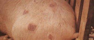
The main distinguishing signs of swine fever are fever, mass mortality, hemorrhagic conjunctivitis, pregnant sows abort hemorrhages in the skin and subcutaneous tissue, as well as on the mucous membranes of the renal pelvis, ureters, bladder, under the epi- and endocardium, under the pleura and mucous membrane of the larynx . Along the edges of the spleen there are black-red infarcts the size of a small nut and dark red staining of the lymph nodes, with a marble pattern on the section, the presence of leukopenia, and in the mucous membrane of the large intestine the presence of characteristic plague buds.
THS is an acute disease mainly in piglets in the first days of life. The causative agent is an RNA virus. The morbidity and mortality rate of viral gastroenteritis in piglets under 10 days of age can reach 100%. At 25 days of age, 15% of piglets die. Adult animals easily tolerate the disease, which is manifested by increased body temperature, diarrhea and agalactia in sows. The disease is characterized by a short-term increase in body temperature by 2-2.5″C. catarrhal-hemorrhagic gastroenteritis, vomiting, diarrhea, dehydration of the body. The incubation period is from 12 hours to 3 days. By the end of 3-5 days, after the onset of the disease, the body temperature drops below normal, which indicates the imminent death of the animal. Treatment is usually ineffective. Porcine rotavirus enteritis is a contagious disease characterized by symptoms of acute enteritis. The causative agent is an RNA virus from the genus Rotavirus of the Reoviridae family. The disease mainly affects piglets 1 - 8 weeks of age and 3-7 days after weaning. Vomiting after feeding is one of the early clinical signs of rotavirus diarrhea. The disease is characterized by profuse diarrhea with subsequent death of 6-50% of piglets. In the timely diagnosis of the disease, isolation of the virus and examination of stool by enzyme immunoassay are of great importance. Passive immunization of weaned piglets with colostrum from cows immune to bovine rotavirus provides a preventive effect.
Intestinal spirochetosis (Spirochetosis of the colon). The disease manifests itself as typhlocolitis and digestive dysfunction. The incidence of disease in a herd can range from 2-30%. The death of animals is rare. The cause of the disease is the hemolytically weakened anaerobic spirochete Brachyspira pilosicoli. It has a morphology characteristic of a spirochete, and its appearance is similar to other species of the genus Brachyspira. The main route of infection is nutritional.
The severity of clinical signs of intestinal spirochetosis is usually noted in the post-weaning period and less often in young animals when switching to a new type of feeding; the disease can also be observed in fattening pigs and from time to time in pregnant sows. The discovery of animals with sunken sides is the first clinical sign of the disease. As a rule, weaned piglets and young animals are born. Affected pigs are depressed, the coat is lustrous and disheveled, the perineum is contaminated with feces, and fever is observed at times. The stools are watery or mucus-like. sticks well to the floor of the machines. The color of the feces is green or brown. Sometimes they contain lumps of mucus and... less commonly, blood clots.
Brachyspira pilosicoli grows well under the same anaerobic conditions described for Brachyspira hyodysenteriae. When infected with an isolated culture with a weak degree of p - hemolysis. 7-8 week old piglets develop mucoid (mucus-like) diarrhea with blood clots and lesions of the large intestine.
Recent advances in improving diagnosis include the use of dual PCR to simultaneously detect Brachyspira hyodysenteriae and Brachyspira pilosicoli.
Proliferative enteropathies. These diseases are also known as porcine intestinal adenomatosis. terminal yelitis, necrotic enteritis, proliferative hemorrhagic enteropathy (or hemorrhages in the digestive canal) [1]. Porcine proliferative enteropathy (PE) is the most common and most frequently diagnosed intestinal infection. The highest incidence is usually observed among adult pigs.
The cause of proliferative enteropathies is an obligate intracellular bacterium - Lawsonia intracellulars, which develops inside the cytoplasm of intestinal epithelial cells.
Salmonellosis often complicates the course of dysentery, especially during the weaning period. The disease is not recorded among adult pigs. Salmonella choleraesuis is more often isolated from sick and dead animals. In this case, the presence of blood in feces is one of the main indicators of the associative manifestation of the disease. A striking feature of the clinical manifestation of salmonellosis is an increase in body temperature to 41-42 C. cyanosis of the skin of the ears, chest, abdomen, inner thighs and damage to the gastrointestinal tract throughout. At the same time, the lungs, liver, and kidneys are affected, and pinpoint hemorrhages are found on the spleen. The mucous membrane of the small intestine is catarrhally inflamed, sometimes with pinpoint or spotty hemorrhages. Changes in the large intestine are more pronounced and are characterized by thickening and folding of the intestinal wall, the development of necrosis of lymphatic follicles and diphtheritic inflammation of the mucous membrane with the presence of dirty gray films on its surface. The infection is easily diagnosed bacteriologically.
Anaerobic dysentery. It is characterized by the appearance of diarrhea mixed with blood and high mortality in piglets aged 1 to 3 days. The causative agent of the disease is Clostridium perfringens type B., multiplying in the intestines, forms a necrotoxin. causing an inflammatory reaction and necrosis in the intestinal wall, which leads to intoxication. The incubation period is short and is determined by hours. Piglets become ill 6-12 hours after birth.
The main symptom of the disease is diarrhea. Sick piglets suckle poorly, their body temperature rises to 410 C. If left untreated, general weakness quickly increases and the animals die within 1-4 hours. At autopsy, the intestine is hemorrhagically inflamed, dark red in color and filled with bloody contents, and necrotic areas are additionally found in the mucous membrane of the jejunum
Timely administration of bivalent serum against infectious enterotoxemia of sheep and dysentery of lambs in combination with antibiotics can be effective.
Enterotoxemia of piglets. The fundamental clinical picture of the disease is severe toxemia after reproduction of Clostridium perfringens in the wall of the jejunum. most often type C. Maximum mortality is observed among piglets 3 - 6 weeks of age. In the case of a hyperacute course, the clinical picture of the disease does not have time to develop, since the sick piglets die after 1-2 days. If the disease continues for several days, in addition to an increase in body temperature, the animals experience anorexia. foamy stool mixed with blood and foul odor.
In adult pigs the disease progresses at lightning speed. Progressive paralysis develops, muscle cramps appear. Death occurs two days after the first signs appear.
Clinical signs of balantidiasis depend on the intensity of the invasion, but they are absent in adult animals. Unlike swine dysentery, the acute course of balantidia is accompanied by an increase in body temperature by 1-1.50 C. Vomiting appears at times. The subacute and chronic course of the disease is manifested by a perverted appetite; body temperature remains normal or increases slightly; piglets lose weight, become weak, and anemic
Trichocephalosis of pigs. The disease is caused by the nematode Trichocephalus suis. In unfavorable farms, the extent of invasion can reach 80% with an intensity of up to 6 thousand or more. Piglets - weanlings and fattening pigs - are more often affected. Animals become infected by eating feed contaminated with infective eggs. The pathogenic effect of whipworms manifests itself in injury to the mucous membrane of the large intestine. By introducing the head end into the intestinal mucosa, the parasites reach the submucosal and muscular layers, causing capillary bleeding.
Weak intensity of invasion is not manifested by pronounced symptoms of the disease. Intensive invasion is accompanied by diarrhea, sometimes with blood and mucus, a depressed state, decreased and perverted appetite, and a frequent increase in body temperature by 1-1 50 C. At times, epileptic seizures with signs of asthma appear.
Esophagostomosis (Nodular disease). The disease is characterized by damage to the large intestines and is accompanied by refusal of food, profuse diarrhea, sometimes mixed with blood and mucus, as a result of which the animals quickly become exhausted. The causative agents of esophagostomiasis in pigs are the helminths Oesophagostomum dentatum and Oesophagostomum longicaudatum belonging to the family Trichonematidaem. The increase in invasion is directly proportional to the age of the animals. The intensity and extent of invasion among young animals 2–4 months of age are insignificant. These indicators progress with age. In adult pigs, the extent of invasion reaches 90%. Animals are mainly infected in walking areas and, to a lesser extent, in pigsties, since esophagostoma larvae are sensitive to the action of decomposing urine.
Clinical manifestations of eimeriosis are recorded in piglets from 10 days to 5 months of age. Adult pigs are parasite carriers.
Pigs carrying Eimeria can be identified both in prosperous and non-prosperous farms. At the same time, eimeria oocysts are found in 82-95% of animals. In some cases, infection of pigs with eimeriosis can reach 100% Symptoms of the disease depend on the intensity of invasion and the general resistance of the animal's body Mortality in the acute course of the disease reaches 100% Subacute course of the disease is often accompanied by conjunctivitis, disruption of the cardiovascular system, encephalitis and pneumonia, decreased or lack of appetite Body temperature rises, defecation becomes more frequent, fecal matter is liquefied with a small amount of mucus and blood.
In conclusion, it should be noted that a number of diseases accompanied by digestive dysfunction can occur with a clinical picture similar to swine dysentery. At the same time, dysentery often occurs in association with other infections, which means that the course of the disease and pathological changes can vary. The main reason for the transformation of the pathological picture of the disease is other intestinal pathogens. Therefore, in every doubtful case, epizootological examinations are carried out. clinical, pathological and laboratory studies of pathological material in order to exclude plague, viral transmissible gastroenteritis, rotavirus enteritis of pigs, intestinal spirochetosis of proliferative enteropathies. salmonellosis. anaerobic dysentery and enterotoxemia in piglets, balantidiasis. trichocephalosis. esophagostomosis. eimeriosis and feed toxicosis.
As a result, differential diagnosis of swine dysentery becomes essential for the correct selection of therapeutic drugs and targeted implementation of health measures
Bibliography
- Diseases of Swine / edited by BE Straw. JJ Zimmerman. S. D'Allaire. DJ Taylor (9th edition). Blackwell Publishing. The Iowa State University Press. Ames Iowa. USA - 2006. - P. 785-799
- Hutto DL and Wannemuehler MJ A comparison of the morphological effects of Serpulina hyodysenteriae or its beta-hemolysin on the murine mucosa / DL Hutto and MJ Wannemuehler// Vet Pathol- 1999. - Vol 36. - P. 412-422.
- Joens LA. Location of Bowden CA and synergistic anaerobic bacteria in colonic lesions on gnotobiotic pigs / LA Joens. RD Glock. SC Whipp I M. Robinson DL Harris // Vet. Microbiol. - 1981. - Vol 6- P. 69-77.
Essay
The article discusses the issues of etiology, pathogenesis and differential diagnosis of swine dysentery. Also, this work outlines the features of the clinical manifestation and transformation of pathological changes in the associative course of the disease with intestinal pathologies of pigs. In addition, information is provided on the isolated manifestation of a number of diseases of a viral bacterial and parasitic nature that occur with clinic for digestive disorders.
Key words: swine dysentery. Brachyspira hyodysenteriae. Brachyspira pilosicoli Brachyspira innocens Lawsonia intracellulans. B-hemolysis
UDC 619:616 9.636.4
SWINE DYSENTERY AND ITS DIFFERENTIAL DIAGNOSIS Buhanov VD, Soldatenko NA, Skvortsov VN
Summary
The questions of etiology, pathogeny and differential diagnostics of swine dysentery are considered in the article. Also, specialties of clinical manifestation and transformation of pathoanatomical changes in the associative course of the disease with intestinal pathologies of pigs are expounded in this work. In addition data on isolated manifestation of number of diseases of viral bacterial and parasitic nature, proceeding with the clinic features of gastroenteric disorder (dyspepsia) are added in the article.
Key words: swine dysentery Brachyspira hyodysenteriae. Brachyspira pilosicoli. Brachyspira innocens. Lawsonia intracellularis. P-haemolysis
About the authors
Soldatenko Nikolai A. chief of laboratory of mycology and mycotoxicology of North Caucasian Zonal Scientific-Research Veterinary Institute: Rostov highway Novocherkassk. 346421. phone: 8 6352 669-92 e-mail
Skvortsov Vladimir N.. D.Sc. in Veterinary Medicine, head of the Belgorod Department of the AII-Russian Institute of Experimental Veterinary. 4. Kurskayast. Belgorod. Russia. 308002: phone/fax: + 7 (4722)26-29-75: e-mail [email protected] ru.
Responsible for correspondence with the editorial board:
Buhanov Vladimir D., Ph.D. In Veterinary Medicine, docent, chief staff scientist of the Belgorod Department of the All-Russian Institute of Experimental Veterinary. Business address: 4, Kurskaya st, Belgorod, Russia, 308002; phone/fax e-mall Home address: 64/7, Narodnyl blvd, Belgorod, Russia, 308001: phone.
Information about authors
Soldatenko Nikolay Aleksandrovich, head of the laboratory of mycology and mycotoxicology, State Scientific Institution SKZNIVI. 346421 Novocherkassk. Rostov highway. GNU SKZNIVI Russian Agricultural Academy. tel: (86352) 669-92 e-mail
Skvortsov Vladimir Nikolaevich Doctor of Veterinary Sciences, Head of the Belgorod Department of the All-Russian Institute of Experimental Veterinary Medicine. 308002. Belgorod, Kurskaya street 4: tel. e-mail
Responsible for correspondence with the editors : Bukhanov Vladimir Dmitrievich, candidate of veterinary sciences, associate professor, leading researcher of the Belgorod department of the All-Russian Institute of Experimental Veterinary Medicine: work address: 308002, Belgorod, st. Kurskaya 4; tel.; e-mail: [email protected] ru; home address: 308001, Belgorod, Narodny Boulevard, 64. apt. 7. Tel. 27-89-85.
| useful links | |||
Treatment of swine dysentery
Treatment of swine dysentery should be carried out comprehensively. Therapy involves several areas, including the following.
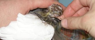
- Eliminate dehydration
. For this purpose, the animal is offered clean water (often with a medicinal product diluted in it, for example an antibiotic solution). If the pig continues to vomit, injectable antiemetics are prescribed. - Reducing the load on the digestive organs
. In the first days from the onset of the disease, the pig is put on a starvation diet, and as the animal’s condition improves, easily digestible feed is gradually introduced into the diet. Before feeding a sick pig, food should be thoroughly chopped and heat-treated - raw food is excluded for the entire period of treatment. - Elimination of infection
. For this purpose, antibacterial drugs are prescribed to the sick animal. The difficulty in treating dysentery is that its symptoms are similar to the clinical manifestations of some other intestinal diseases. But the rapid onset and rapid development of the disease often does not leave time for diagnosis and identification of the pathogen. This makes it reasonable and advisable to use broad-spectrum antibacterial drugs that are active against most pathogens of infectious gastrointestinal diseases in pigs.
Such drugs include Tylosin AVZ. Its active ingredient is tylosin, an antibiotic from the macrolide group, which exhibits high activity against most bacteria, chlamydia and mycoplasmas. Its effect appears within 1–3 hours after application, and the duration of influence reaches 18 hours.
"Tylosin AVZ" is fed with water, which is convenient both for the owner of several heads and for large pig farms.
Veterinary
19.11.2013
THIAMULIN-CONTAINING DRUGS FOR THE TREATMENT OF SWINE DYSENTERY
Porcine dysentery
(Disenteria suum) is an infectious disease characterized by hemorrhagic diarrhea and catarrhal-hemorrhagic inflammation of the large intestine with necrotic changes in the mucous membrane. The causative agent of this disease is a spirochete from the Treponematidae family, which is classified in the 1st group of infectious disease agents in terms of resistance to disinfectants. In the external environment at a temperature of about 0°C, the pathogen can persist for up to 9 weeks, and at temperatures up to 20°C - up to 2 weeks. The incubation period for Brachyspira hyodysenteriae ranges from 7 to 60 days.
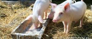
Pigs of all breeds and age and sex groups are susceptible to the pathogen, but especially young animals up to 5–6 months. A clinically recovered animal continues to release the pathogen into the external environment for another 90 days. The disease is recorded at any time of the year, but less often in the summer and during camp conditions. The source of the pathogen is sick and recovered animal carriers, and the reservoir of infection is mice and rats.
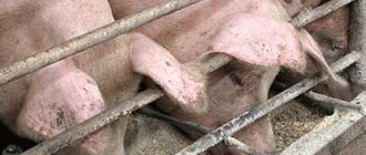
Spread of swine dysentery
contribute to: overcrowding, unsanitary conditions, hypothermia, high levels of bacterial carriage, lack of vitamins in the diet, feeding with low-quality feed, illness from other infectious diseases (influenza, pasteurellosis, etc.). Infection of animals occurs through the nutritional route through food and water, care items, transport and other objects contaminated with the secretions of sick animals. It should be taken into account that piglets, especially in winter, can get sick several times until they die or reach 5-6 months of age. The economic damage from dysentery consists of the death of animals, reaching 30–40% or more among weaned pigs, up to 10% in adult pigs, low feed conversion, weight loss and loss of breeding value of recovered animals.
The main symptom of swine dysentery
- diarrhea with the presence of mucus or mucus with blood in the stool. The incubation period of the disease is 10–15 days. It occurs acutely, subacutely and chronically.
The acute course lasts 2–7 days, body temperature is normal or elevated (can reach 41°C), due to incessant diarrhea, animals quickly lose weight and die. The corpses are cachetic, the tail and area of the hind limbs are stained with liquid, foul-smelling brown feces. An autopsy reveals catarrhal or catarrhal-hemorrhagic inflammation of the intestine.
In the subacute course - debilitating bloody diarrhea, emaciation, weakness, tightness of the abdominal wall with retraction of the sides. Piglets also often die.
The chronic course of the disease can drag on for up to 1–3 months; if there is an appetite, the animals are severely stunted in growth. The feces are liquid or soft, sometimes with blood or mucus.
For the treatment of swine dysentery
At different times, various chemotherapeutic agents and regimens of use are recommended: use of a drug based on tylosin intramuscularly at a dose of 10 mg DV/kg body weight once a day, 3–5 days; use of a drug based on metronidazole intramuscularly at a dose of 5–10 mg DV/kg body weight or orally with drinking water at a dose of 75–100 mg/1 l of water for 3 days. However, all these methods and means do not allow achieving a complete cure of the entire affected population; therefore, it is necessary to find new effective and easy-to-use therapeutic agents.
One of the most effective drugs of choice for the treatment of swine dysentery is the drug “Tialong” produced by Nita-Pharm CJSC. Tialong as an active ingredient contains tiamulin hydrogen fumarate (in terms of tiamulin base) at a concentration of 100 mg/ml. Tiamulin, the active ingredient, was discovered during a study of the antimicrobial activity of a close relative of the oyster mushroom, and further experiments confirmed its effectiveness and safety. Tiamulin is more soluble in fats than macrolides and even lincosamides (eg lincomycin). Since the cells lining the intestines and blood vessels, as well as bacterial organisms and membranes, are approximately 80% fat or lipid, therefore, tiamulin is absorbed to a greater extent from the intestines and penetrates deeper into bacterial cells more than any of the macrolides. Macrolides have valuable biological properties that distinguish them from antibiotics of other groups: high activity against spirochetes, mycoplasmas and gram-positive microorganisms, including those resistant to penicillins and cephalosporins, good tolerability when administered, high degree of bioavailability, low toxicity to the macroorganism and lack of cross-resistance with antibiotics of other groups.
For swine dysentery, Tialong is used at a dose of 100 mg DV/12.5 kg body weight (1.0 ml/12.5 kg body weight) intramuscularly once; if necessary, according to indications, the injection can be repeated after 24 hours.
Extensive production tests carried out under production conditions confirmed the high therapeutic and prophylactic effectiveness of the drug “Tialong” for swine dysentery.
Further introduction of the drug "Tialong" into veterinary practice will make it possible to take a significant step in solving the problem of dysentery in pig farming. Number of impressions: 5809