Chickens are one of the most unpretentious poultry. With proper care, birds can be raised all year round. A thrifty owner will always have fresh dietary meat and eggs on his table. But for year-round rearing, it is important to know the peculiarities of bird development, how long the development of a chicken lasts, as well as the subtleties of the intimate life of birds, the technology of incubating eggs and keeping young animals.
Chicken development
Interesting facts about the chicken and the egg
First, a few fun facts that will be of interest to poultry farmers:
- Chicken eggs can be artificial. They learned to make these in China. The shell is made from calcium carbonate, and the inside is made from gelatin, aromatic additives and dyes. Externally, the product is practically no different from the natural one, but its taste is different, and there are practically no beneficial properties.
- There are people who are terrified of eggs. This fear has a scientific name - ovophobia. The oval shape causes panic. Every thousand people on Earth suffer from mental illness.
- In South Sulawesi, a breed of roosters was bred that, instead of crowing, makes sounds similar to human laughter. Feathered gulls bring decent profits to breeders.
Cackling rooster from South Sulawesi
- There are several yolks in an egg. Double-yolk fruits are often produced by very young or very old hens. Sometimes three, four or even five yolks are hidden under the shell. A larger number has not yet been registered.
- The color of the shell does not affect the quality of the contents. Thus, white, brown and variegated eggs contain approximately equal amounts of valuable elements. However, a dark color indicates a denser shell, which means that such eggs stay fresh longer.
It is worth noting that the size and weight of the egg depends on the breed, age and body characteristics of the laying hen. Occasionally, even an average chicken can “give out” a product weighing up to 150 grams when the norm is 55–75 grams. But there are those that produce tiny eggs, comparable in size to quail eggs.
Even an ordinary chicken can lay a giant egg
How long does it take for a chicken to develop?
Hatching chicks is called incubation. It can be natural or artificial. This period lasts an average of 21 days. Then the chickens mature and with proper care, by the 49th day, broilers reach 1.5 kilos of live weight. A two-month-old broiler chicken weighs an average of 2 kg. Egg breeds weigh slightly less.
Young roosters are distinguished by their comb, beard and spurs.
At 2 months, roosters show the first signs of puberty, hens are a month late. In young males, the comb, beard and spurs become noticeable, the legs lengthen and become more massive. Rounded feathers grow on the tails - braids. But these signs do not mean that they can mate and produce healthy offspring. Roosters are ready for fertilization at the age of 24 weeks.
The hen usually lays her first egg at 22 weeks.
The indicator of the onset of oviposition and readiness for reproduction in females largely depends on the breed. On average it is 22 weeks. But some hens produce their first eggs as early as 17 weeks. When birds of different breeds begin to fly:
- birds of complex productivity and hybrids - at about 6 months;
- fighting - after 9 months;
- light, small species - at 4.5–5 months.
If a chicken goes through puberty in the autumn-winter period, it will start laying eggs later than expected. The feathered lady managed to “mature” before the cold weather, and in winter she will live in a warm poultry house? She will supply eggs even on frosty days, but, of course, in smaller quantities. Thus, a pattern can be identified: females from winter broods begin laying eggs first, and females from summer broods begin laying eggs later.
The number of eggs laid depends on the breed and age
How many eggs can you get per day? On average one. In a year, you can get about 2 hundred eggs from a bird of the egg breed; meat breeds give about 1.5 hundred. Egg production also depends on the age of the chicken. Most bird breeds are maximally fertile in the first two years of life. Then egg production decreases, and by the age of five the bird becomes useless. The number of eggs does not depend on the presence of a rooster. The male is needed only for breeding offspring.
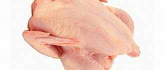
Eggs from broiler chickens
This type of chicken is bred specifically to obtain meat products from these individuals in a short time. In this case, representatives of meat (Cornish) and meat-eating (Plymouthrock) breeds are taken as a basis. During the breeding process, the best characteristics of the maternal and paternal lines are selected.
Those obtained as a result of such selection have a unique precocity - by 1.5 months, chicks with a well-designed diet can gain up to 1.5 kg, and already at 2 months the young weigh up to 5 kg.
But subsequently, the chickens’ weight gain decreases sharply, so their further keeping becomes unprofitable. So usually broilers are fattened until they are two months old and then simply slaughtered.
Raising broilers to produce egg products is difficult even on an industrial scale in poultry farms where there are specialists. And on personal farmsteads it is useless to keep such hybrids as layers - broilers are bred solely for the sake of producing tasty, tender meat.
To obtain egg products, chickens for egg and meat-egg production should be bred.
Physiology of broiler chickens
And the physiological structure of broiler chickens is such that if their females begin to lay eggs, then they begin to have health problems and end with their death. And although Russian breeders are working on this problem and are trying to breed crosses that, in addition to meat, will also lay eggs, there has not been much progress in this direction yet.
But broiler chickens can still lay eggs; their sexual maturity occurs approximately at the same time as other breeds - from six months to 8 months. But some hens can start laying eggs only when they are one year old.
Interesting! Do broilers lay eggs and how do they reproduce? Genetically, it so happens that broiler hens lay only double-yolk eggs.
How does a rooster impregnate a hen?
One rooster should have an average of 10 girlfriends
The sexual physiological characteristics of the rooster are not visible outwardly; the intimate organs of birds of both sexes are the same and represent a cloaca. In roosters, the testicular canals exit into the cloaca, and in chickens, the oviduct. During sexual intercourse, the cloaca come into contact and the seed enters the hen's oviduct. Fertilization takes place in the oviduct funnel - sperm penetrate there after mating. They can be stored there for up to 20 days: the eggs will be fertilized during this time, even if the rooster does not trample this hen.
On average, there should be 10–16 females per male. Heavy beef breeds may have a ratio of 1:6.
A small harem will lead to the fact that a loving rooster will simply torture the females. They will look plucked and unhealthy, because in a fit of passion the male sits on the lady’s back and grabs the scruff of the neck with his beak.
Rooster tramples hen
However, it is worth noting: the fewer coitus a rooster has per day, the more “live” eggs there will be, and, accordingly, offspring. In large farms, where one male is not enough, roosters of various age categories are kept. Otherwise, bloody battles for leadership cannot be avoided.
Video - A breeding rooster impregnates a hen
Incubation of chicken eggs: determining the optimal mode
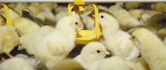
Day old chicks.
Source: Wikimedia When incubating chicken eggs, one of the most important conditions for obtaining high-quality young animals is the use of biologically complete, fertilized eggs for incubation and maintaining optimal incubation conditions - temperature and air humidity.
To assess the quality of the starting material - hatching eggs - biological control methods are used. Biological control is carried out both before incubation and throughout it, and even after incubation.
High-quality hatching eggs are obtained from healthy birds with adequate feeding and optimal housing conditions.
Before incubation, chicken eggs are carefully selected, sorted and evaluated according to the following parameters:
- appearance (purity and quality of the shell, uniformity in shape and size);
- egg mass;
- mobility of the yolk and its color (when transilluminated);
- the state of the white and yolk and their color (when opening the eggs).
Cancellation of chicken eggs using an ovoscope is the main method of biological control used during the incubation process. The eggs are examined using an ovoscope in order to determine the correct development of the chicken embryo (embryo); the number of unfertilized eggs and dead embryos. The ovoscope allows the poultry farmer to determine the quality of eggs and select the optimal incubation mode.
After incubation, the number of healthy young animals hatched, their growth (especially in the first weeks of rearing) and safety are taken into account. Chicks are assessed at one day of age based on their appearance.
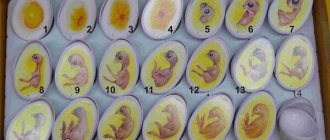
Table of chicken embryo development during incubation
How to track the process of embryonic development in an egg? To do this, you need to view eggs (for example, chicken) on an ovoscope, open several eggs with normally developed embryos at different stages (for example, 3–4, 6–7, 10–11, 18–19 days) and compare them with photographs of embryos of the corresponding age .
There are many embryonic eggs in the ovary of birds. During the breeding season, they grow, accumulating nutrients. After maturation, the egg enters the oviduct, in the upper section of which fertilization occurs. Then the egg (yolk) moves along the oviduct, gradually becoming enveloped in protein and two subshell membranes. A shell is formed in the lower part of the oviduct. Thus, in birds, embryonic development begins while the egg is passing through the oviduct.
Bird eggs are extremely rich in nutrients, which are abundantly sufficient for the growth and development of the embryo. A freshly laid egg contains a germinal disc or blastodisc. When such an egg is opened, it is easy to distinguish a small round spot on the yolk with a light central part (light field) and darker edges (dark field).
After laying, the egg cools and the development of the embryo stops. He falls into a latent state, remains alive, but his metabolism is reduced to a minimum. Such a break in development can last painlessly for 5–7 days; with longer periods, the embryo weakens and dies.
During storage, eggs lose moisture, some of it passes from the white to the yolk, carbon dioxide is released, and the pH of the white and yolk increases. The physical properties of the shell also change - its surface becomes shiny. Due to the evaporation of moisture, spotting (“marbling”) occurs.
The process of chick formation
Formation of a chicken in an egg by day
First, eggs develop in the ovary (their number sometimes reaches three thousand). As it matures, each egg with a supply of nutrients, that is, the yolk, is sent to the oviduct, where the protein is formed. If mating has occurred, she will be fertilized. But without fertilization, the egg will also mature, acquiring a shell at the last stage - in the uterus, attached to the cloaca. The latter reaches 60 cm in length in adult hens, and 20 cm in pullets.
Egg maturation lasts from 23 to 26 hours. Unfertilized products are no different in taste and beneficial properties from fertilized ones.
The development of the chick begins with the blastodisc - a tiny clot of cytoplasm on the surface of the yolk. It is formed in the laying hen’s body, and then, in the egg, cell division continues.
This is what a blastodisc looks like on the yolk
After a couple of days of incubation, the development of the amnion and allantois begins. These are temporary organs that ensure the vital functions of the future chicken. The amnion protects against mechanical injury and drying out - it is responsible for the regulation of fluid depending on the age of the embryo. Allantois supplies oxygen, removes waste products, and supplies valuable substances and minerals from the shell.
Development of the allantois and its closure
The outer structure of the shell is called the cuticle. It prevents pathogenic bacteria from entering. Eggs should not be rubbed or washed, otherwise this protection will disappear and they will spoil.
In the first days of embryo formation, valuable substances from protein and yolk are nourished. This is enough to provide all the initial needs of the embryo. But by the middle of the development period, a large embryo is located along the egg with its head towards the blunt tip. The protein from the sharp end is displaced and, with the help of the amnion, enters the embryo's mouth.
Chicks gain most of their weight in the first two months after hatching.
Part of the yolk in the later stages of development will be drawn into the abdominal cavity and will become the baby’s food in the first days after hatching. Then the fluffy will begin to peck soft food: chopped eggs, cottage cheese, chopped greens. From two weeks of age, babies are offered a mixture of different types of flour, and from one month of age - coarse grains. The greatest weight gain occurs in the first couple of months after birth: in egg breed chicks it increases by 16 times, in meat breeds it increases by more than 30 times.
Video - Development of a chicken egg by day
Histology.RU
Material taken from the site www.hystology.ru
Sex cells. Fertilization . Splitting up. The sperm of birds, like those of other representatives of vertebrates, have a flagellated shape and consist of a head, neck, and tail.
Rice. 55. Scheme of the structure of a chicken egg:
1 - shell; 2 - latebra; 3 - subshell membrane; 4 - white yolk; 5 - yellow yolk; 6 - Pander's nucleus (white yolk lying under the latebra); 7 - blastoderm; 8 - vitelline membrane; 9 — air chamber; 10 - egg white (outer layer of albumin); 11 - egg white (fibrous layer); 12 - chalaza; 13 - chalaza-like layer; 14 - egg white (inner layer of albumin). The curve on the right shows the rate of egg growth during the 18 days leading up to its laying. The dashed lines leading from the different layers of the yolk to the growth curve show exactly when these layers were formed.
In different species of farm birds they differ in size, shape of the head and tail. For example, the upper part of the sperm head of a rooster is blade-shaped, while in a gander it is elongated and pointed at the end.
The lifespan of sperm in the female genital tract is more than 30 days. During this period, without re-mating, hens lay fertilized eggs. In the first two weeks after mating, the number of fertilized eggs is greatest.
Based on the number and location of the yolk, bird eggs are classified as polylecithal or telolecithal. A bird's egg contains: the yolk, that is, the egg, the white, the subshell membrane and the shell (Fig. 55). All components of an egg, except the yolk, belong to the tertiary shells. Their material is produced by the glands of the oviduct. As the egg moves through the oviduct, it becomes covered with tertiary membranes.
The egg, like other cells, has a nucleus and cytoplasm. The nucleus, located at the animal pole of the cell, is surrounded by a thin layer of cytoplasm containing organelles. The rest of the egg is filled with yolk, a cellular inclusion. The yolk contains proteins and lipids. As a result of concentration from a dissolved state, they precipitate and take the form of balls. In the initial stage, the yolk globules are rich in phospholipids and then in saturated fatty acids. Chickens have a lot of free cholesterol in their yolk balls.
The yolk of an egg is not uniform; it can be light or dark. In the center of the egg there is a light yolk. This zone is called latebra and is shaped like a flask. In the rest of the egg, the dark and light yolk are arranged in layers. Typically, the light yolk is deposited at night, and the dark yolk at other times of the day.
The peripheral layer of the cytoplasm of the egg is called the cortical layer, and the membrane of the egg, or plasmolemma, is called the primary membrane. In the white, the egg is suspended on chalazae, directed from the yolk to the blunt and sharp end of the egg. Chalazas are bundles of thin twisted fibers consisting of a dense protein-carbohydrate complex.
The chalazae hold the yolk in the center of the egg so that the embryo is always on top.
Outside the yolk is the white. Based on the location of its location, the outer and inner layers are distinguished, each of which consists of liquid and dense protein. Egg white contains water (87%), proteins, glycoproteins, free carbohydrates, a small amount of lipids, ash and other substances. For the developing embryo, protein is the main source of nutrients and water during the middle period of embryogenesis. Protein is a supplier of minerals and proteins. It contains bactericidal substances that kill microorganisms.
The protein is covered with a subshell membrane, in which two layers are distinguished: outer and inner. The outer layer, about 56 microns thick, consists of fibers with a diameter of 2 - 10 microns. This layer is tightly connected to the shell. The thickness of the inner layer is 16 microns, and the diameter of its fibers is 2 - 3 microns. The chemical composition of the fibers is similar to a keratin-like (horny) substance. The inner layer of the subshell membrane is adjacent to the protein.
In the area of the blunt end of the egg, the layers of the subshell membrane diverge and an air chamber containing air is formed.
The subshell shell is very dense, elastic and permeable to gases, water, and soluble compounds. The properties of the shell largely depend on its moisture content. The moist subshell membrane swells, the pores in it increase, the role of which is played by the spaces between the fibers.
The contents of the egg are enclosed in a shell. The latter protects the egg from damage; its material is used by the embryo as minerals in the construction of the skeleton. The shell has pores through which gas exchange and evaporation of moisture occur during the development of the embryo.
The shell contains organic and mineral substances. Densely woven fibers and protein granules are built from organic substances. The outside of the shell is covered with a thin film above the shell - cuticle. Constructed from mucin, it prevents the penetration of microorganisms and fungal spores through the pores of the shell and is permeable to gases.
After leaving the ovary, the egg enters the oviduct, where it is fertilized. When a male mates with a female, several million sperm enter the oviduct at the same time. A feature of fertilization of eggs in farm birds is polyspermy - up to 300 sperm can penetrate into the egg, and only one sperm connects to the nucleus of the female reproductive cell. After fertilization, moving along the oviduct and becoming covered with tertiary membranes (albumen, subshell membrane, shell), the egg intensively divides by mitosis. Therefore, laying an egg is an embryo at an early stage of embryogenesis.
Cleavage in birds is meroblastic (partial) or discoidal. Only the animal pole of the zygote takes part in it, where the nucleus and cytoplasm, devoid of yolk, are located. The vegetative pole does not participate in crushing, since it is loaded with yolk, which inhibits crushing.
The first two crushing furrows have a meridional direction and are located perpendicular to each other. Meridional furrows give way to latitudinal ones (Fig. 56). The cells (blastomeres) formed during crushing lie on the yolk in the form of one layer - a disk, therefore the crushing is called discoidal, and the resulting blastula is called a discoblastula. Its roof and marginal zone consist of disc blastomeres, and its bottom consists of undivided yolk.
The blastocoel has the appearance of a narrow slit and is shifted towards the animal pole of the blastula (Fig. 57).
Rice. 56. Crushing a chicken egg:
A - 2 blastomeres; B - 4 blastomeres; B - 8 blastomeres; G - about 16 blastomeres.
Rice. 57. Germ disc of a chicken egg at the late stage of cleavage (A) and at a stage close to gastrulation (B).
1 - blastomeres; 2 - ectoderm; 3 - blastocoel; 4 - yolk.
After fertilization, the egg can remain in the oviduct for 4–27 hours, so both cleavage and gastrulation occur in this organ.
In the laid egg, embryogenesis stops and resumes again from the moment of incubation or incubation.
Gastrulation . Laying of the axial organs. Development of germ layers. After 12 hours of incubation, a germinal shield forms in the center of the germinal disc. From this part of the germinal disc the body of the embryo develops. The rest of the disc is called non-embryonic, since it is used in the construction of fetal membranes (temporary, or provisional, organs) that ensure the normal development of the chick. The embryonic shield is surrounded by a light field. Its cells are raised above the yolk. Below them is a subembryonic cavity formed as a result of the embryo using the yolk. The light field is surrounded by a dark field. Its cells rapidly divide and grow over the surface of the yolk. This zone is called the fouling edge.
Gastrulation in birds, as in other vertebrates, occurs in two phases. First phase: by stratification, or delamination, of a single-layer disc, two germ layers are formed - ectoderm and endoderm (Fig. 58). The second phase is the formation of the chordomesodermal rudiment: the cells of the marginal zone of the disco-blastula intensively divide and migrate towards the posterior edge of the embryonic shield in two streams, where they meet
Rice. 58. Gastrulation in birds at an early stage. Delamination and emigration:
1 - ectoderm; 2 - endoderm.
and begin to move forward along the midline of the germinal shield, forming a thickened cell ridge - the primitive streak. It is directed from the posterior edge of the embryonic shield forward.
In the middle zone of the primary stripe, a depression is formed - the primary groove (Fig. 59), at the anterior end of which a thickening appears. This is the primary, or Hensen's, node. It has a depression called the primary fossa (gastric sac).
The primitive streak is identical to the blastopore of the gastrula of lancelets and amphibians. The primary fossa of Hensen's node is a homologue of the dorsal lip of the blastopore, the remaining zones of the primary streak are homologous to the lateral and ventral lips.
In the zone of the primary fossa, the embryonic material invaginates and, in the form of a cellular cord, moves between the ectoderm and endoderm to the anterior end of the embryo. This cord is called the head, or chordal, process, from which the notochord develops (Fig. 60).
Two wing-shaped primordia migrate through the primary groove towards the anterior end of the embryo. They grow on the sides of the notochord between the ectoderm and endoderm.
Rice. 59. Gastrulation in birds:
A - migration of cells in the germinal shield; B — formation of chordomesodermal primordium; 1 - ectoderm; 2 - material of the future neural plate; 3 — material of the notochordal plate; 4 - primary (head) nodule; 5 - primary fossa; 6 - primary stripe; 7 - primary groove; 8 - chord; 9 - mesoderm. Solid arrows indicate the direction of movement of material in the outer germ layer, and dotted arrows - in the middle germ layer (according to Knorre).
Rice. 60. Changes in the germinal shield during gastrulation (A):
1 - primary streak formed by mesodermal material; 2 - primary (head) nodule formed by the rudimentary material of the notochord; 3 - rudiment of the neural plate; B - four transverse sections of the gastrula, made at the levels indicated in Figure A (a - through the anterior end of the body, b, c - through the middle section of the body, d - primary nodule); 1 - cutaneous ectoderm; 2 - neural plate; 3 - mesoderm; 4 - chord; 5 - intestinal endoderm.
The differentiation of these primordia ends with the formation of the middle germ layer, the mesoderm.
Thus, in birds, gastrulation ends with the laying of the germ layers. In the gastrulation of birds, a further complication in the development of vertebrates is noted: the rudiment of the notochord and mesoderm completely lost contact with the primitive gut. The latter is very early separated from other embryonic primordia and is part of the endoderm of the embryonic shield.
Further development of the germ layers in birds proceeds as in other vertebrates (see Fig. 53). The neural plate is released from the ectoderm, and after joining its edges, the neural tube is formed. The ectoderm creeps from the outside onto the neural tube, and therefore the latter becomes submerged under the ectoderm. The neural tube is the source of development of the entire nervous system, and the ectoderm is the rudiment of the surface layer of the skin (epidermis).
The mesoderm is divided into somites (segmented mesoderm), segmented legs, and unsegmented mesoderm (splanchnotome). The somites include the dermotome, myotome, and sclerotome. The deep layers of the skin develop from the dermotome, the muscle tissue of the skeleton develops from the myotome, and the skeleton from the sclerotome. Segmental legs are the source of development of the urinary system.
In the splanchnotome there are parietal (external) and
Rice. 61. Formation of axial primordia in a chicken embryo:
A, B, C - three successive stages; 1 - neural groove; 2 - neural tube; 3 - ectoderm; 4 - chord; 5 - mesoderm; 6 - somites; 7 - visceral leaf of the splanchnotome; 8 — parietal leaf of the splanchnotome; 9 - intestinal endoderm.
visceral (internal) layers, between which there is a coelom - the secondary body cavity. Epithelial tissue develops from the splanchpotome, covering the serous membranes of the internal organs, the thoracic and abdominal cavities (Fig. 61).
Formation of extraembryonic organs. The development of the embryo's body is accompanied by the formation of temporary, or extra-embryonic (provisional) organs that help create the necessary conditions for the normal development of the embryo. Provisional organs in birds include: yolk sac, amnion, serosa, allantois. They are also built from germ layers - ectoderm, endoderm, sheets of unsegmented mesoderm. The source of development of these leaves is the extraembryonic zone of the germinal disc (color table I).
The rate of development of temporary organs can often outstrip the development of the embryo; in birds, compared to other animals, this process occurs more intensively.
The formation of provisional organs begins with the growth of extra-embryonic material of the blastodisc over the surface of the yolk of the peripheral zone. This zone is called the fouling zone, and after gastrulation is complete, it consists of extraembryonic ectoderm, endoderm, and sheets of unsegmented mesoderm.
Subsequently, during the development of the fetal membranes, the ectoderm is always associated with the parietal layer, and the endoderm is always associated with the visceral layer of mesoderm.
Before other membranes, the yolk sac is formed, which is formed in the process of fouling of the yolk with germ layers. It performs a trophic function and is built from extraembryonic endoderm and visceral layer of mesoderm. The endoderm of the yolk sac is involved in the enzymatic breakdown of the yolk and the absorption of nutrients into the blood vessels located in its mesodermal layer. Since the bird egg contains a large amount of yolk, this membrane is very well developed. The yolk sac is laced from the body of the embryo by the trunk fold. Its formation is due to the intensive proliferation of cells of all germ layers lying on the border of the embryonic and non-embryonic material. All germ layers of this zone participate in the trunk fold. The tip of the fold is directed under the body of the embryo, and therefore the embryo rises above the yolk, and its trunk endoderm, previously spread out on the yolk, forms the intestinal tube. The latter communicates with the yolk through a narrow vitelline duct.
The trunk fold initially develops in the head part of the embryo and then spreads in the caudal direction.
Amnion (amnion - cup), or water membrane. Serosa . Simultaneously with the trunk fold, which separates the embryonic from the non-embryonic part, the amniotic fold is formed. Its crest is directed above the body of the embryo. Appearing first above the head of the embryo, it gradually engulfs the body. The head part of the amniotic fold forms very quickly, so its wall consists only of ectoderm. After fusion of the edges of the head part of the amniotic fold, proamnion appears. The trunk zone of the amniotic fold lags behind in development. It consists not only of ectoderm, but also of the parietal layer of mesoderm.
Above the body of the embryo, the edges of the amniotic fold grow together: ectoderm with ectoderm, parietal layer with parietal layer of mesoderm. After the edges of the amniotic fold have fused, the body of the embryo is surrounded by two membranes—the amnion and the serosa.
The formation of the amnion involves the ectoderm and the parietal layer of the mesoderm of the inner half of the amniotic fold, and the second - the ectoderm and the parietal layer of the outer half of the amniotic fold. The edges of the caudal part of the amniotic fold do not fuse together; a canal is formed here that connects the amnion cavity with the rest of the egg. This channel is called the gray-amniotic channel. The ectoderm of the amnion produces amniotic fluid, which fills the amniotic cavity, creating the most favorable aquatic environment for the developing embryo. The amnion performs a protective function, it smoothes out shocks and creates the possibility of some mobility for the embryo.
The serosa is the very surface membrane of the embryo. Adjacent to the shell, it covers the outside of the amnion with the embryo located in it and the yolk sac with the yolk.
The main functions of the serosa are participation in gas exchange and the transfer of calcium ions from the shell to the body of the embryo. The electron microscopic structure of serosa ectoderm cells is similar to cells that produce chlorides: the plasmalemma is equipped with numerous microvilli, and there are a lot of mitochondria in the cytoplasm. These cells synthesize hydrochloric acid, the action of which is apparently aimed at dissolving the calcium salts of the shell, which are then transferred to the fetus.
Allantois (allantois - sausage-shaped) is formed after the completion of intestinal formation on the ventral surface of the hindgut. It, like the intestine, consists of endoderm and visceral mesoderm. The allantois gradually fills all the gaps between the amnion, yolk sac and serosa, merging with the mesodermal layer of the latter. A dense network of blood vessels is formed in this area, which helps improve the oxygen supply to the body.
Thus, adjacent to the shell, the allantois participates in gas exchange. It performs the function of an excretory organ, since metabolic products accumulate in it.
All provisional organs are reduced at the end of embryogenesis. From the above material it follows that the development of birds also occurs in stages. Disruption of the typical course of one of the stages entails changes in the entire course of development of the embryo. Knowledge of the stages of development allows us to identify the most important periods in the formation of the embryo. Understanding them is extremely necessary in connection with the tasks of practical poultry farming.
However, at present, periodization in the development of birds is not yet sufficiently developed. The development of the chick embryo has been studied in most detail.
H.P. Tretyakov and M.D. Popov, based on the nature of the embryo’s nutrition, developed the following classification of stages of chicken development.
Stage 1 - latebral nutrition - lasts for the first 30 - 36 hours. The material for nutrition is the yolk of the latebra, containing proteins, salts, and water. The source of energy is glycogen, so the embryo requires almost no oxygen. There is no blood circulation in the body.
The 2nd stage - yolk nutrition and the presence of the yolk circulation - lasts from 30 - 36 hours to 7 - 8 days of incubation. By the 30th hour of incubation, the heart and yolk sac are formed, and blood vessels develop in the latter. The glycogen supply almost completely disappears. Oxygen enters the body, which facilitates the use of proteins and fats. Other membranes, as well as body organs, are formed: the nervous system develops, the heart begins to beat and the muscles of the body begin to contract. The formation of the liver determines the beginning of urea synthesis, so the breakdown products of proteins become less harmful to the embryo.
The 3rd stage - breathing oxygen from the air and feeding on egg white - lasts from the 7th - 8th day to the 18th - 19th day. By the end of it, the embryo becomes a formed fetus. This stage is characterized by intensive development and functioning of the allantois, in the wall of which a dense network of blood vessels is formed. Adjacent to the serosa and with it to the subshell membrane of the egg, the allantois provides oxygen supply to developing tissues. Thanks to the dissolution of the shell, the intensity of mineral metabolism increases and the absorption of fats increases.
Stage 4 - consumption of oxygen from the air chamber of the egg. The chicken switches to pulmonary breathing, the pulmonary circulation begins to function, and arterial blood appears, enriching the tissues with oxygen. The stage lasts from the 18th - 19th day until hatching. At this point, the allantois undergoes reverse development, and the chick begins to lack oxygen. It pecks at the inner leaf of the shell membrane and begins to breathe the air contained in the air chamber of the egg.
The 5th stage - hatching - lasts from the 20th to the 21st day. The chicken feeds on the remains of the yolk, which, together with the yolk sac, are drawn into the intestinal cavity. Other embryonic membranes also stop functioning and die. The chicken pecks at the shell and leaves the egg.
There are other classifications. According to the periodization developed by G. A. Schmidt and M. H. Rogozina, based on the embryogenesis of the chick embryo, four periods of development are distinguished: embryonic, prefertal, fetal, and also the hatching period.
The embryonic period lasts the first eight days, of which the initial stages of development occur during the movement of the embryo through the oviduct. The embryonic period is characterized by intensively occurring processes of morphogenesis. The source of nutrition for the embryo is the yolk and the liquid fraction of the protein partially mixed with it. Once the yolk sac is formed, nutrients are transported through its blood vessels. Respiration of the embryo first proceeds osmotically, and then is provided by the vascular system of the yolk sac. At the end of the embryonic period, the blood vessels of the allantois take part in this process.
The prefetal period begins at the end of the 8th day and ends by the 13th day of incubation. All provisional organs reach functional maturity. Along with the main extra-intestinal method of feeding the embryo, which is carried out through the yolk sac, the material is absorbed intra-intestinal: the chicken swallows amniotic fluid from the amnion cavity.
Respiration of the prefetus occurs exclusively through the vascular system of the allantois.
The fetal period covers the period from 13 - 14 days to the beginning of the 20th day of incubation. Characteristic of this period is the intensive growth of the organs of the fetal body. The main method of feeding the embryo is intraintestinal; the extraintestinal method becomes additional. The protein that moves into the amnion cavity and mixes with the amniotic fluid is a source of nutrition. Allantoic respiration is preserved. The blood vessels of the allantois are located near the subshell membrane. This helps to enhance gas exchange, which is necessary due to the intensive development of the chicken’s body organs.
The hatching period is on the 20th - 21st day of incubation. It is characterized by cessation of the functions of temporary organs, intestinal digestion, and breathing with the participation of the lungs.
Thus, data on the adaptive reactions of the body and the patterns of morphogenesis characteristic of each period of embryogenesis provide a scientific basis for the development and implementation of measures aimed at further improving the technology of artificial breeding of farm birds.
Reviews (0)
Add a review
Selection of eggs for hatching and incubation
Not all “fruits of the labors” of hens are suitable for hatching. The selection must be done extremely carefully.
How to select chicken eggs for hatching?
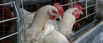
Step 1: Focus on the age and health of the hens. Chickens of egg breeds should not be younger than 7 months, meat breeds - 8 months. They should be checked for developmental pathologies or infectious diseases (colibacillosis, pullorosis), because the infection can be transmitted to offspring.
It is necessary to decide on the breed of chickens and check the health of the laying hen
Step 2: Select eggs that are the right shape and size. Deformed ones can indicate pathological diseases in the laying hen. Too large or small ones are also not suitable. Ideal size: for egg breeds it is 50–60 grams, for meat breeds – 50–70 grams, and for meat breeds – 55–70 grams.
The right eggs to hatch
Step 3: Inspect the shell. It needs to be clean, without chips, nicks, or growths. In addition, it should not be thin, porous or marble-like in texture. This indicates a lack of minerals.
For hatching you need eggs with a smooth shell without blemishes.
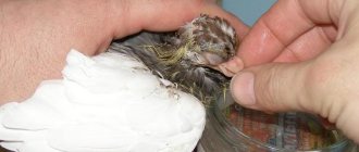
Step 4. Check with an ovoscope to see if the eggs are fertilized. You can purchase the device or make it yourself from a box with a hole on the wall that matches the transverse diameter of the egg, and a lamp with a power of 100 W or more. Ovoscopy is performed in a darkened room. Hold each egg close to the hole and hold it up to the light. In the center of the fertilized egg there will be a uniformly colored yolk with a dark spot of the embryo. Unfertilized ones and those with more than one yolk are discarded.
Checking an egg with an ovoscope
Step 5. During ovoscopy, pay attention not only to the yolk, but also to the air compartment - the chamber needed for the embryo to breathe. It is located near the shell in the blunt part. If the chamber is moved, the egg should be discarded.
This egg has a breathing chamber in place
Do not forget that it is better to take fresh eggs for hatching, no older than four days. If you need to collect a large number of them, the period can be extended to 7 days. At a storage temperature of +15°C and a humidity of about 75%, the embryo will survive.
Additional check with an ovoscope
An ovoscope is used to check the eggs during the formation of the embryo. This is done three times:
- On the 6th day, when the circulatory system should appear. If the yolk field is uniform or only a blood ring is visible, then the embryo is dead.
- On the 10th day. A noticeable blood network appears and the allantois closes. If this is not the case, and the embryo looks like a dark spot, the egg should be removed.
- On the 18th day. The outlines of the embryo become noticeable, and the neck at the blunt end trembles.
Pecking begins on the 19th–20th day, depending on the breed (earlier in eggs), and a day later the chick is born.
The babies can be born in 20 days
Prices for ovoscope
Ovoscope
What is “ovoscoping”
An ovoscope is a device that helps select eggs for laying and further cultivation in an incubator (photo). Such artificial brooding methods are necessary for breeding chicken breeds that have poorly developed maternal instincts. These include most decorative breeds, “Hungarian Giants”, “Holoshecks” and others.
Ovoscope
The ovoscope shines a beam of bright light through the egg, which allows you to see what is happening inside the shell throughout all stages of development. Constant candling helps to control the process and timely weed out eggs unsuitable for breeding.
Important! X-raying with an ovoscope is performed no more often than once every 3 days.
The ovoscope allows you to track all stages and stages of chicken development in the egg and identify pathologies such as:
- immobility of the yolk when rocking;
- failure of the yolk to return to its place after shaking;
- marbling of the shell;
- two yolks (such an egg cannot hatch two chickens);
- light or dark stripes;
- lack of yolk;
- dark spots and light stripes;
- absence of embryo inside.
Candling eggs
The following factors indicate normal chicken development:
- clearly visible vascular network;
- an embryo entangled with vessels is visible;
- It is noticeable how the chicken moves at the very end of the maturation period.
Natural way to raise chickens
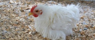
The best hens are made from birds of heavy breeds. Some egg-bearing females have virtually no maternal instinct. And large feathered ladies are more diligent. But before placing carefully selected eggs under the hen, it is necessary to determine whether she is ready for “motherhood.”
Large quoits are more assiduous
In a female ready for incubation:
- body temperature increases;
- the comb becomes red and fades slightly;
- the belly increases, and the number of feathers on it decreases.
Chickens from eggs
The main phases of development of a chick in an egg
Have you ever wondered how the embryo develops inside a chicken egg? Let's shed some light on this seemingly everyday, yet magical process.
The embryo develops inside the egg in two stages. The first passes inside the chicken’s reproductive tract, the second - outside its body, in the external environment.
During mating, the male's sperm enter the funnel through the oviduct, where fertilization of the egg occurs. After this, fragmentation of the germinal disc begins.
The entire process of development of the embryo in the oviduct occurs within 24-27 hours, at a mother’s body temperature of 40.5-41 ° C, under conditions that completely exclude the evaporation of water. When an egg enters the external environment, it begins to cool, water evaporates from it and development slows down.